

 English
 Français

Welcome to the Academy
Here you will find all the resources you need to train, develop your ICU diary skills and find inspiration!
Quick access
Straight to the point
Explore the app
I want the patients' point of view
Ready to use
What should I actually write?
I want to go further
Find all the definitions
FAQ
Who and why?
Who should have an ICU diary?
For any patient in the following situations:
• Cognitive impairment for more than 48 hours
• Coma
• Sedation
• Agitation
• Delirium
• Conscious but unable to communicate
• Any patient who wants one
When should an ICU diary be opened?
• As early as possible, as soon as it is foreseeable that the patient will spend more than 48 hours sedated, in a coma or with delirium
• From the first day of unconsciousness or delirium
=> Save time on the diary's opening message
=> Limit the loss of information about the first hours/days
What are the contraindications?
• There are no absolute contraindications
• Relative contraindications include:
- language barrier (but translation software can be used)
- low digital literacy (but the diary can be printed using LifeMapp Diary's export function)
Who can open an ICU diary?
•Any clinician caring for the patient
My patient is conscious and doing fairly well, but I know we will need to intubate them later (transplant plan, foreseeable deterioration). When should I open their diary?
• I can open their ICU diary immediately
• With patient access, they can use all the application's features, including managing relatives, and personally invite the people they want to write in their diary. They can also assign them to the desired privacy circles.
• This is an ideal situation!
Do I need to obtain the patient's consent before opening an ICU diary?
• In most cases, it will not be possible to ask for the patient's consent, as they are rarely in a position to give informed consent.
• As with any other aspect of care, relatives should be informed and, where applicable, the trusted person should be asked for agreement.
Do I need to obtain relatives' consent before opening an ICU diary?
• As with any other aspect of care, relatives should be informed and, where applicable, the trusted person should be asked for agreement.
Is the patient's consent obtained at another point?
• When the ICU diary is created in LifeMapp Diary, if the patient's email address is known and entered, an email is sent so that they can create an account and access their diary. That email includes a button allowing them to delete their information and access without logging into the platform, thereby enabling retrospective consent, following the model used in research involving patients who are unable to be informed at the time.
Note: this email is sent again when the patient leaves intensive care and can also be resent by clinicians and the diary's relative administrator after discharge from intensive care.
What is an ICU diary for?
• Provide the patient with a written, chronological record of their stay in intensive care
• Help them fill in the 'ICU memory gap', the amnesia
• Help them reclaim their story
• Help them make connections between delirious memories and real events from their stay in intensive care
• Help them better understand their relatives' experience
Are ICU diaries recommended?
• Keeping an ICU diary has been included since 2023 in the French National Authority for Health (HAS) recommendations on preventing Post-Intensive Care Syndrome, and has also been recommended by intensive care scientific societies since 2009.
Are ICU diaries new?
• ICU diaries have been used in intensive care since the 1980s. Originally written only by nurses, they are now shared between relatives and clinicians.
Open an ICU diary
How do I open an ICU diary with LifeMapp Diary?
• Click “Create a diary” on the Home page
• Enter the requested information
• Confirm creation of the diary
I don't have my patient's email address. What should I do?
• Above all, do not enter anything else in the “patient email” field
• You can complete creation of the ICU diary without any problem: simply skip the question and come back to it later.
• Do your best to obtain the patient's email address later.
• As long as no email address has been entered, you will receive a reminder each time the diary is opened and when the patient's diary is closed.
• If no email address is ever entered, the patient will never be able to use LifeMapp Diary with their own access.
• If the patient does not have an email address, you can export the ICU diary as a PDF when the patient leaves, print it and hand it to them directly.
Can I enter someone else's email address (for example, a spouse's) in the patient email field?
• NO. This would breach the confidentiality of the system.
• WARNING: the patient's email address is the address to which their personal access details will be sent and will become their username for logging in to LifeMapp Diary.
• Only patient access provides access to all messages, including private messages.
• It is essential to not to enter another person's email address here, to avoid any breach of confidentiality.
• 101 accepts no liability for any problem of this kind caused by incorrectly entering the patient's email address.
My patient shares an email address with their spouse/partner. What should I do?
• Enter the shared email address in the “Lead relative” section.
• Do not enter anything in the patient email field.
• You can then choose to:
• Print the ICU diary for the patient (possible with clinician-administrator access)
• Create an individual email address for the patient when they leave
• Add the shared address to the patient field as well, but only with the patient's direct, informed agreement after waking. This means the spouse/partner may be able to access every message in the diary.
I obtained my patient's email address after creating the diary. What should I do?
• In the diary list, click “Manage”, then “Edit diary information”.
• Scroll down, click “Edit diary information” and enter the email address in the patient field.
• After entering the address, go back to “Manage”, then select “Resend invitation to patient”.
I don't have the lead relative's email address. What should I do?
• You can complete creation of the ICU diary without any problem: simply skip the question and come back to it later.
I obtained the lead relative's email address. What should I do?
• In the diary list, click “Manage”, then “Edit diary information”.
• Scroll down, click “Edit diary information” and enter the email address in the patient field.
• After entering the address, go back to “Manage”, then select “Resend invitation to patient”.
What happens when I create an ICU diary?
• If you entered the patient and/or lead relative email addresses, an email is sent with a link allowing each person to create an account.
• Otherwise, nothing happens until you add those email addresses.
What does the patient receive when their ICU diary is created?
• If you entered the patient's email address, an email is sent with a link allowing them to create an account.
• That email includes a few explanations about the principles of the ICU diary.
• The same email will be sent again automatically when the patient leaves.
What does the lead relative receive when an ICU diary is created?
• If you entered the lead relative's email address, an email is sent with a link allowing them to create an account.
• The same email will be sent again automatically when the patient leaves.
Write in an ICU diary
Who can write in an ICU diary?
• All clinicians caring for the patient, whatever their profession or grade, have something to contribute to the ICU diary. It is important that this care is delivered as a team!
• Each profession can describe its relationship with the patient and the care provided within its own field of practice.
• The patient's relatives who have been approved by the lead relative (or, where applicable, the trusted person)
Can several people write together?
• This is even recommended at the beginning, when you are not yet very confident, so that nothing is forgotten, to feel reassured about what is written, or to build skills.
• Remember to sign the message together at the end!
Are any types of content prohibited in an ICU diary?
• An ICU diary is not a medical record or a repetition of clinical handovers. Examination results or detailed diagnostic, therapeutic or prognostic information should not be written in it.
Note: the patient has the right to request a copy of their medical record from the institution. The ICU diary is not intended to duplicate that document.
• An ICU diary must respect professional confidentiality while informing the patient (after the event) and their relatives, as required by law:
« In the event of a serious diagnosis or prognosis, medical confidentiality does not prevent the family, the patient’s close relatives or the person of trust defined in Article L.1111-6 from receiving the information they need to provide direct support, unless the patient objects” French Public Health Code, Article L 1110-4
« In the event of a serious diagnosis or prognosis, medical confidentiality does not prevent the family, the patient’s close relatives or the person of trust defined in Article L.1111-6 from receiving the information they need to provide direct support, unless the patient objects” French Public Health Code, Article L 1110-4
• If you need to write information that you do not want to share with the whole family but only with the closest relatives, you can tick “Sensitive content” at the end of your message in LifeMapp Diary.
=> “Sensitive content” in LifeMapp Diary is accessible only to the patient, clinicians and the diary's relative administrators — no one else. Other relatives do not have access.
What should I write in an ICU diary as a clinician?
• Writing in an ICU diary means taking the patient's point of view and telling them about events in terms of their lived experience and the care relationship.
• Ask yourself: looking back on this day from the patient's point of view, what will be important to tell them?
• What might leave traces — on their body or in their memory?
• Everyday events during the hospital stay:
• Daily activities: washing, small care procedures, mobilisation, etc.
• Environment: anything the patient can perceive — light, sounds, etc.
• Clarify anything that might otherwise be misinterpreted
• Patient reactions
• Movements
• Agitation, delirium
• Pain
• Relaxation, etc.
• Daily activities:
• Visits from relatives
• Procedures
=> in terms of anything that may leave memory or physical traces: scars, sensations
• Examinations/tests
=> as an event or trip out of the room, not the details of the indication or result
• Key milestones during the stay:
• Progress
• Awakening
• First milestones
• Deteriorations
How should I start an ICU diary?
• For the patient, it is important that the story starts at the beginning: tell them about their admission, what happened during the first hours, and recap all the major events at the start of the stay.
• You can add a photo of the empty room, either showing the whole room or taken from the patient's point of view by standing at the head of the bed, so that they later have a visual record of that perspective.
Should I write on days when difficult things happen? (deterioration, cardiac arrest, etc.)
• YES! It is part of the patient's intensive care story.
•The ICU diary should help patients reconstruct the reality of their stay in intensive care. Content should respect readers' sensitivity without sugar-coating reality.
• Difficult moments should also appear so that patients can understand why rehabilitation takes time.
• Patients often look in their diary for major moments: the date of the cardiac arrest as well as the date they opened their eyes again.
• You do not have to describe the resuscitation manoeuvres in detail if that makes you uncomfortable. You can simply write: “You worried us a lot today; your condition deteriorated.” The patient can ask questions afterwards if they feel the need.
Should I write on days when nothing happens?
• YES! From the patient's point of view, there is never really “nothing” happening in intensive care. All the small things are interesting:
• Describe how a shampoo is done for an intubated patient
• Explain that temperature is measured in the ear, to make sense of the cold sensation in the ear that may leave strange memories
• Mention the flickering fluorescent light, a baby crying in the next room, anything the patient may have perceived…
=> A gap in the diary raises questions: nothing on that date — did something serious happen?
Who can read what I write?
• The patient and members of your team can access all your messages.
• For relatives, LifeMapp Diary has three privacy circles:
• Relative administrators have access to everything except private messages.
• First-circle relatives can access non-sensitive, non-private messages from relatives and clinicians.
• Second-circle relatives can access only non-sensitive, non-private messages from relatives.
What are “privacy circles”?
• LifeMapp Diary allows the patient and relative administrators to manage the other relatives added to the ICU diary.
• The lead relative (entered when the diary is created) or the first relative administrator can appoint up to three additional relative administrators (maximum four per diary). Relative administrators assign the other relatives to the other two circles and can access everything except private messages they did not write themselves.
• The first circle is intended for relatives who would visit the patient in intensive care (or have a similarly close level of intimacy). They can access messages from clinicians and relatives, except messages labelled “Sensitive content” and “Private messages” that they did not write themselves.
• The second circle is intended for relatives who are less closely connected to the patient. They can write messages of support and can access only non-sensitive, non-private messages from relatives that they did not write themselves.
As a clinician, can I move someone to a different privacy circle?
• No. Only the relative administrator(s) and the patient can do this. This protects you: you cannot interfere in family management, and you cannot be blamed for it. Responsibility lies with the relative administrator(s).
What are “private messages”?
• Private messages are a feature available only to relatives and the patient.
• Only the patient and the person who writes the private message can access it.
• This preserves a degree of privacy within the ICU diary.
• This can be useful in some complex family situations.
• Note: clinician administrators and the patient can export the diary with or without private messages.
• In the PDF export with private messages; these messages are marked with a small padlock
•Warning when exporting a PDF WITH private messages (a feature available only to patient accounts and clinician administrator accounts), you are responsible if it is not handed directly to the patient and a relative comes into possession of messages they were not meant to read.
What is “sensitive content”?
• Sensitive content is a feature available to everyone (patients, relatives and clinicians): anyone can mark their own message as sensitive.
• Sensitive messages can only be opened by:
• The person who wrote it
•The patient
• Clinicians
• The diary's relative administrators
• This feature is useful for limiting access to certain messages to these people.
• Note: clinician administrators and the patient can export the diary with or without sensitive content.
• In the PDF export with with sensitive content, these messages are marked with a crossed-out eye icon
• Warning: when exporting a PDF WITH sensitive content, a feature available to clinician-administrator accounts, you are responsible for handing it directly to the patient. If a relative gains access to messages they were not meant to read, responsibility lies with you.
Is LifeMapp Diary secure?
•LifeMapp Diary is fully secure: the application complies with GDPR principles; data is hosted by OVHcloud in France on HDS-certified health-data infrastructure; the invitation system uses a unique token that prevents unauthorised access to diaries and unit platforms; and an SSL certificate protects entered information through encryption.
Manage my team
Healthcare staff are divided into two categories: administrator staff and “standard” staff. The rights assigned to each category are as follows:
✅ indicates a permission ❌ indicates that this category does not have access | Create an ICU diary | Write in an ICU diary | Create a new clinician account | Export ICU diaries as PDF (excluding private messages) | Patient transfer | Access to the unit monitoring dashboard |
Clinician administrators | ✅ | ✅ | ✅ | ✅ | ✅ | ✅ |
Non-administrator clinicians | ✅ | ✅ | ❌ | ❌ | ✅ | ❌ |
Recommendations for project success
• Identify a core group of healthcare staff motivated by the project, who will form the group of clinician administrators for the service platform
• Ideally, they should come from the different teams and professions represented in the unit.
• They should have attended the training.
• They are responsible for asking every day: “Have we remembered to open an ICU diary for this patient?”
• They should be identified by the rest of the team as a resource in case of a problem or question.
• They are responsible for monitoring the unit dashboard and contacting 101 if the project slows down.
• They are responsible for regularly updating the list of clinician user accounts for the unit: sending invitations to new arrivals, regularly checking that clinicians who have left the unit have deleted their account and, if not, deleting them themselves.
• They liaise with the 101 team to report problems, questions and suggestions, and to request a refresher webinar or training when needed.
• Make access and reminders easier
• Install shortcuts to the LifeMapp Diary login page on the unit's computers (bedside computers, unit workstations, offices, etc.).
• Add a shortcut to the LifeMapp Diary login page within the clinical software.
• Add a daily or twice-daily task (per shift/team) to the end-of-shift task list or checklist for patients who may benefit from an ICU diary.
• Use reminders in the form that best suits your organisation: computer wallpapers, posters in patient rooms or care stations, etc.
• Engage the team
• Organise a symbolic reward for clinicians with the highest number of messages each month.
Support relatives
How do I give my patient's relatives access to the ICU diary?
• A first relative administrator must be appointed:
• Either when the ICU diary is created, by completing the fields in the “Lead relative” section
• Or later: go back to the ICU diary information using “Edit diary information” and click “Invite relatives” once you are inside the diary.
• Without a first relative administrator, no other relative can be invited.
• This first relative administrator will then be responsible for accepting or rejecting the other relatives.
• You can invite any relatives you meet in the patient's room. We recommend doing so without ticking “Relative administrator”, so that you do not take on responsibility for family access management.
• When you follow this recommendation:
• The relative administrator receives a notification that another relative is waiting for approval.
• They can accept or reject that person.
• When the person is accepted, the circle-selection screen appears, reminding the administrator of each circle's permissions, and the relative administrator must assign the person to the appropriate circle.
What can relatives write in the ICU diary?
• Any moment shared with the patient during their intensive care stay
• Visits
• Activities during visits
• Patient reactions
• Etc.
• What is happening in life around them:
• Life at home without them
• Family events
• Relevant news (politics, sport, etc.)
• The changing seasons
• Without going into excessively intense emotions: relatives' experiences, difficulties and worries
• And of course support, affection, tenderness, plans for the future and encouragement
A relative complains about a clinician or the care provided in the ICU diary. What should I do?
• Occasionally, relatives may misuse the ICU diary by treating it as a way of communicating with clinicians.
• Respond quickly by contacting the person who wrote the complaint in the ICU diary.
• First of all, make sure the complaint is heard: let the person explain the problem, acknowledge the emotion, and redirect them to the appropriate channel so it can be addressed — the unit's management team, the hospital's patient-relations department, etc., depending on the context and issue.
• Once the complaint has been heard and acknowledged, remind them that the ICU diary is not a communication tool for the team or a complaints book:
• Clinicians are not required to read the messages, so the complaint may receive no response.
• The ICU diary is intended for the patient: when you write in it, you are addressing the patient and only the patient.
A relative is expressing a lot of emotion in the ICU diary. Is that a problem?
• This can happen, especially when only one relative is writing: the ICU diary can become a private journal in which they pour out an overflow of emotion.
• For the patient, an ICU diary that is too emotionally intense can be very difficult to read: they may feel like a voyeur when reading highly intimate writing, or feel guilty for having caused so much suffering to their relatives.
• It is useful to start a conversation with the relative and remind them that the ICU diary is intended for the patient.
• Do not hesitate to suggest that the relative keep a separate private journal alongside the ICU diary, where they can express all those emotions. Writing can indeed be cathartic, and it can be very helpful to have a place to let them out.
• Suggest that the relative imagine the moment when the patient reads it: will they feel comfortable with the patient reading those words?
What are “private messages”?
• Private messages are a feature available only to relatives and the patient.
• Only the patient and the person who writes the private message can access it.
• This preserves a degree of privacy within the ICU diary.
•This can be useful in some complex family situations.
• Note: clinician administrators and the patient can export the diary with or without private messages.
• In the PDF export with private messages; these messages are marked with a small padlock
• Warning: when exporting a PDF WITH private messages, a feature available to clinician-administrator accounts, you are responsible for handing it directly to the patient. If a relative gains access to messages they were not meant to read, responsibility lies with you.
What is “sensitive content”?
• Sensitive content is a feature available to everyone (patients, relatives and clinicians): anyone can mark their own message as sensitive.
• Sensitive messages can only be opened by:
• The person who wrote it
• The patient
• Clinicians
• The diary's relative administrators
• This feature is useful for limiting access to certain messages to these people.
• Note: clinician administrators and the patient can export the diary with or without sensitive content.
• In the PDF export with with sensitive content, these messages are marked with a crossed-out eye icon
• Warning: when exporting a PDF WITH sensitive content, a feature available to clinician-administrator accounts, you are responsible for handing it directly to the patient. If a relative gains access to messages they were not meant to read, responsibility lies with you.
What are “privacy circles”?
• LifeMapp Diary allows the patient and relative administrators to manage the other relatives added to the ICU diary.
• The lead relative (entered when the diary is created) or the first relative administrator can appoint up to three additional relative administrators (maximum four per diary). Relative administrators assign the other relatives to the other two circles and can access everything except private messages they did not write themselves.
• The first circle is intended for relatives who would visit the patient in intensive care (or have a similarly close level of intimacy). They can access messages from clinicians and relatives, except messages labelled “Sensitive content” and “Private messages” that they did not write themselves.
• The second circle is intended for relatives who are less closely connected to the patient. They can write messages of support and can access only non-sensitive, non-private messages from relatives that they did not write themselves.
As a clinician, can I move someone to a different privacy circle?
• No. Only the relative administrator(s) and the patient can do this. This protects you: you cannot interfere in family management, and you cannot be blamed for it. Responsibility lies with the relative administrator(s).
Take photos
Can I add photos of the patient?
• Yes. It is even recommended if the patient's relatives think the patient would be interested in seeing them.
• Studies and patient interviews now show that many patients appreciate having photos of themselves taken at different stages of their stay: to keep a record, reclaim their image and see what others saw. Many patients regret not having any, having too few, or not having images of the “good moments” with clinicians or visitors.
• However, this is not the case for 100% of patients, so it is always useful to ask relatives for their opinion, as they know the person and can guide you.
• If relatives think it is not a good idea, you can simply take photos from the patient's point of view or of the environment without including the patient's face.
• If you add a photo of the patient:
• Prefer moments when the patient is comfortably positioned and has company.
• Always add explanatory text to the photo, stating that it was taken for the patient to help them reclaim their image, and explaining what can be seen in the photo.
• Always tick “Sensitive content” in LifeMapp Diary. This hides the photo behind a cover that the patient can choose whether or not to open and restricts who can see it.
=> “Sensitive content” in LifeMapp Diary is accessible only to the patient, clinicians and the diary's relative administrators — no one else. Other relatives do not have access.
• If you decide to take photos, remember to do so at regular intervals and at major milestones so that the patient can see their progress.
What other photos should I take for the patient?
• Any contextual photo can be useful!
• Patients often really appreciate photos of their room, both the view they had and other viewpoints that help them understand the space where they spent several weeks — show them what was behind them! They are often very curious about this.
• They also appreciate photos of the team caring for them. If you are comfortable with that, go for it!
• Patients are often curious about what lies outside their room too: show them the waiting room where relatives go during care procedures, the nurses' station where they hear you talking, etc.
• If you take them outside, take photos of that moment too — patients often experience it very intensely.
• All the good moments!
What is the best way to take a photo?
• When LifeMapp Diary is used on a phone or tablet, photos can be taken directly in the application without being stored elsewhere first. We therefore recommend taking photos this way, so that no patient image passes outside LifeMapp Diary or is stored elsewhere.
• LifeMapp Diary is designed to store these photos securely, and stored data is encrypted.
What are the good practices for photos in an ICU diary?
• Photos of the patient should be realistic and reflect reality, but not be “extreme”: take them when the patient is comfortably positioned and covered appropriately to preserve dignity.
• It is useful to include context: avoid a close-up of the patient alone; instead, include a relative or clinician in the photo where possible.
• It is essential to accompany every photo of the patient with an explanatory note stating:
• Why the photo was taken (for example: “Many patients would like to have photos of themselves from their stay in intensive care, which is why we wanted to make this possible for you.”)
• An explanation of everything visible in the photo: machines, equipment such as tubes and catheters, etc.
• It is also important to document good moments and the patient's progress. Some patients regret having only a few photos from the beginning of their stay and no record of the “good memories” of intensive care (yes, they exist!).
My patient is leaving
I want to transfer my patient's ICU diary to another unit
• You can transfer an ICU diary to another service that uses LifeMapp Diary. To do so, in the diary list, click “Manage” and then “Change service”. A window will open where you can select the receiving service.
• Once confirmed, the ICU diary is transferred and disappears from your lists. An automatic message is displayed in the patient's diary indicating the transfer.
• Clinician administrators in the receiving unit receive an email notification that an ICU diary has been transferred to them. It now appears in their list of open diaries.
My patient has left the unit. What should I do?
• When a patient leaves your unit, you are responsible for recording their discharge by closing their diary. To do this, in the list of diaries, click “Manage” and then “Close diary”.
• An automatic message is displayed in the patient's diary indicating discharge from intensive care.
Note: the patient and relatives can continue writing for another six months.
• Once the diary is closed, it moves to the “Discharged” tab in your diary list. It will remain there for six months.
My patient is returning to the unit. What should I do?
• If your patient was discharged less than six months ago, you can reopen their diary by finding it in the Discharged tab of your list, then clicking “Manage” and “Reopen diary”.
• After six months, you need to open a new ICU diary.
Can my patient and their relatives continue writing in the ICU diary?
• Yes. For six months after discharge, the patient and relatives can continue using the application as before. After six months, the ICU diary is archived and can no longer receive new contributions.
What happens in archive mode?
• At the end of the six-month “Discharge” period, the diary is automatically archived. It will remain there for two years, during which the patient and their relatives can continue to access the app to read and download the diary PDF, but can no longer write entries.
• After two years, the archive is permanently deleted.
Who can export the ICU diary?
• Only the patient and clinician administrators can export the complete ICU diary, and they have several export options (with or without private messages, and with or without sensitive content)
• All relatives can export the content they had access to in the application, no more and no less.
• All relatives can export the content they had access to in the application, no more and no less.
My patient has died
• In the event of death, you can close the diary using “death” mode. Nothing particular will happen: no email will be sent and no indicator will appear in the diary. We nevertheless recommend informing relatives before taking any action in LifeMapp Diary.
Reading
*under construction*
General knowledge
*under construction*
I still have a question
Write to us at lifemapp@one-o-one.eu and we’ll get back to you!
Demos
Example bank
Admission
Example #1
Hello Mr Dupont,
You were admitted to intensive care today with very severe breathing difficulties. When you arrived, you were extremely anxious and having great difficulty breathing.
We quickly put you to sleep and placed you on breathing support to ensure your organs received enough oxygen. You seemed calmer when the sedative medication was given.
We also placed an arterial catheter so we can take the regular blood samples needed to monitor your breathing without having to prick you each time. This small catheter was placed in your wrist, below your left thumb. You may have a tiny mark there when you wake up.
Deteriorations
Example #1
Hello Mrs Durand,
This afternoon, things became a little more difficult for your heart. Your blood pressure dropped and we needed to give you a medication called noradrenaline to help your heart and blood vessels maintain a satisfactory blood pressure.
We increased the dose several times and closely monitored your heart and urine output. Your blood pressure gradually improved.
Your family has been informed of what happened and will be arriving soon. They are very upset. We will accompany them into your room and explain again anything they need clarified.
Progress
Example #1
Hello Mrs Lucas,
I'm Aurélie, the night nurse. I was with you when you were intubated last week.
For the first time, I can finally communicate with you. The breathing tube is bothering you a lot, but you need less and less oxygen, and that is really good news.
I can’t wait to hear your voice again. Until then, we’re communicating as best we can… but I think we’re doing pretty well 🙂
See you tonight!
Example #2
Extubated!!!
The whole team is celebrating this major victory.
We know there is still a long road ahead, but this first milestone gives us energy for what comes next.
Another dialysis session today, but it looks like your kidneys are starting to work again.
We're keeping a close eye on things…
I'll be here tomorrow, so I'll let you rest now.
Days when nothing happens
Example #1
Hello Mrs Martin,
You are still sedated and the ventilator is continuing to breathe for you.
At 11 a.m., I helped wash you. After the wash, we rolled you onto one side and then the other so we could change your sheets.
We also gave you mouth care, using small soaked sponges to apply mouthwash inside your mouth.
The ties holding your breathing tube in place were replaced with new ones.
I wish you a lovely day!
Example #2
Hello Mr Dupont
Sometimes, when the intensive care unit is noisy, your eyebrows furrow and your blood pressure rises a little… I wonder whether the environment is making you feel stressed?
There are other patients in the unit, and their machines can make noise… and of course the staff can too!
Maybe you're feeling worried?
Some patients later told us that they heard voices and sounds but found them difficult to understand. The ventilator seemed like a steam engine, or lying on the mattress felt like being on a boat. Did you have this kind of perception?
Example #3
Hello Mrs Durand,
You are still sedated, but we have started reducing the doses. For now, you are still deeply asleep.
With my colleague Jean, we took advantage of having a little time today to give your hair a good wash. It really needed it — your hair was beginning to get tangled. Washing the hair of an intubated patient is always quite an adventure!
We moved you well up in the bed so that your head was right at the top of the mattress, then slid a basin of warm water under your head to wet your hair. You wrinkled your nose a little at first and then relaxed completely. Maybe this will become a pleasant memory between two ICU dreams?
Example #4
Hello Mr Dupont
Last night was particularly hectic. The patient in the room next to yours deteriorated, and we spent a large part of the night in their room caring for them.
You may have heard our voices and all the machine alarms, which we sometimes tried to talk over by raising our voices. The corridor light stayed on so we could move around safely, as did the light in your neighbour's room. The rooms do not block light very well, so you also had light for much of the night.
It is hard to know what you are perceiving around you at the moment; you are still “between two worlds”… when you wake up, you can tell me whether you noticed all this activity.
Have a good day!
Visits
Example #1
Hello Mr Dupont,
Today I'm looking after you together with Charlène, your nurse. We had big plans for you, and together we've managed to carry them out!
This morning Adrienne, the unit physiotherapist, got you doing a little cycling and you tolerated it well, so this afternoon we're planning to sit you out in a chair! That will be a first.
We hope we can get you into the chair before your wife and son arrive, so we can surprise them 🙂
They are here very often; you can feel so much love at every visit. We usually put two chairs on either side of the bed, but today one of them will sit on the edge of the bed and you'll be in the big, throne-like chair!
Delirium
Example #1
Dear Mrs Y,
You are still on the ventilator and sometimes you seem a little confused.
We communicate in writing, and you wrote something about spiders and strange people in your room. At times you seemed frightened.
ICU delirium is quite common and can have different causes. We will try to rule out possible causes and reassess everything.
Keep going — your wife has arrived, and we're all taking good care of you!
Awakening
Example #1
Hello Mr Dupont,
The sedatives were stopped three days ago, and over the last few hours you have started responding more clearly when we ask you to close your eyes and squeeze our hands.
In our jargon we call that “following simple commands” — you must have heard that phrase many times around you! Things like “CLOSE YOUR EYES” or “SQUEEZE MY HAND”, sometimes said rather loudly… It helps us know whether your movements are reflexes or voluntary and lets us assess how you are waking up.
As you can see, waking up is not like it is in the movies, especially after being asleep for a long time. It happens in small steps, and this is a big one! Well done 🙂 I hope to see you even more awake tomorrow.
Discharge
Example #1
Hello Mrs Dupont,
There we are: no more tube, no urinary catheter, normal food again, and I have just removed the central line you had in the left jugular vein, in your neck.
You're going back to the unit you were in before intensive care — at last you'll see daylight again!
I wish you a good recovery and all the best as you return to your new home!
All the best
Death
Example #1
Mr X,
This morning at 6:50 a.m., you passed away surrounded by all your loved ones — family and friends. Your playlist was playing in your room.
Our thoughts are with you and your loved ones.
Goodbye,
Jenny, Laura and Mickaël
Example #2
To the ladies I met several times (partner, sister, mother and little Margot),
I would like to send you my warmest thoughts at this difficult and painful time.
I hope we were able to support you as well as possible.
!! I still need help !!
I would like to submit an anonymised post for feedback: write to us at lifemapp@one-o-one.eu
!! I want to help my colleagues !!
I would like to submit an anonymised post to add to the collection: write to us at lifemapp@one-o-one.eu
LifeMapp Diary resources
Sample protocol to adapt to your needs

PDF ICU diaries
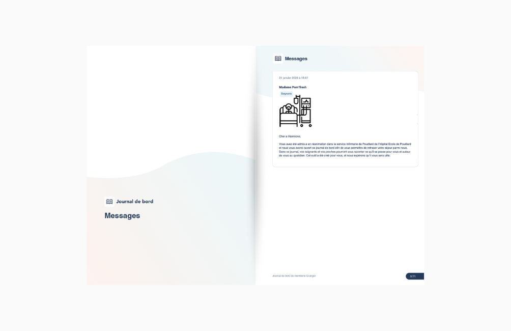
To let you see what an ICU diary looks like when exported as a PDF, the Fonds 101 team has created several fictional diaries that you can freely view.
Business card for our tools
If you would like to have the cards printed by a professional printer, simply send them the print-optimised PDF. The cards are 85 × 55 mm.

LifeMapp Diary flyer
This flyer, intended for relatives, helps them retrace the patient's story during their stay in intensive care.

LifeMapp flyer
This flyer introduces LifeMapp to relatives.

Second Life flyer
This flyer presents Second Life and is intended for patients and their relatives.

Second Life poster
Need Second Life posters for your corridors, waiting rooms, break rooms or changing rooms? You can now download one.

LifeMapp Diary login QR code
You can download the LifeMapp Diary login QR code to display it in patient rooms, on your materials, etc.

Scientific resources
Recommendations
*under construction*
Articles
*under construction*
Glossary entries
View all existing Glossary entries
Adrenaline
An emergency medicine given in very small doses during resuscitation after cardiac arrest, or as a continuous, precisely controlled infusion. It helps the heart work more effectively.
The administration may go completely unnoticed by the patient, especially if they are unconscious, but some describe a feeling of “the human body’s machine getting a boost”, warmth and sometimes headaches.
Enteral feeding
When patients can no longer eat by mouth (because of sedation, coma, swallowing problems, etc.), they can be fed through a tube placed in the stomach or, in some cases, the intestine: a gastric tube. This is called enteral feeding or enteral nutrition (enter- means “stomach”).
Enteral nutrition means feeding the person directly into the stomach or intestine. In intensive care, it is used when the patient cannot eat normally but their digestive system is still working.
The nutrients (proteins, sugars, fats, vitamins, etc.) are given in liquid form to meet the body’s needs and prevent malnutrition.
This method is closest to natural eating and helps keep the intestine working properly.
From the patient’s point of view:
They look like cream-coloured bags of liquid connected by tubing to the gastric tube. The tubing passes through a pump that regulates the flow rate.
You can see the liquid flow from the bag through the tube and into the gastric tube.
If the liquid is at room temperature or chilled, you may feel it flowing slightly through the tube, but it is painless and tasteless. Most of the time, you feel neither hunger nor fullness, as nutrients are delivered at a constant rate throughout the day. In some cases, enteral feeding can be given only at night to try to stimulate hunger during the day and help the digestive tract return to a normal diet.
Parenteral nutrition
In some cases, the patient’s digestive tract may not be working or may not be usable (for example, because of surgery, an infection or a blockage). Nutrients are then delivered directly into the blood through an infusion into a large vein (see Catheter).
This is known as parenteral feeding or nutrition.
In intensive care, this method provides the patient with the energy and proteins needed for recovery, even when the digestive tract is not working.
Most parenteral nutrition comes in bags of white liquid. These products are usually infused through catheters because they can irritate small veins over time.
Amenorrhoea
It is common for women’s periods to become irregular or stop during an intensive care stay or hospitalisation. The body experiences such a shock that it shuts down its reproductive functions to focus on survival.
Amnesia
Amnesia means loss of memories.
Patients often have no memory, or only partial memories, of their time in intensive care. There are many causes of amnesia: sedatives and anaesthetics often affect memory; severe infection and sleep disorders directly affect the ability to form memories; and other mechanisms may also play a part.
Patients who experience amnesia describe a kind of “absence from oneself”. They may have no memory of the events before they were put into a coma, the time spent in a coma, or the period until waking—and sometimes for several days afterwards. “It’s as if someone forgot to put the film in the camera,” one patient said. Many patients feel a need to fill in this “gap” in their story, sometimes soon after leaving intensive care and sometimes several months later.
To support the psychological recovery of intensive care patients, it can be helpful to help them understand what happened during the periods they cannot remember. Keeping a diary is a simple and effective way to do this.
Analgesia
Pain relief, or analgesia, is the removal of the sensation of pain. It can be achieved with medicines (painkillers) or without medicines. Depending on the pain being treated, heat (a hot-water bottle), cold (an ice pack) or physiotherapy can also be very effective.
Anaesthesia
Anaesthesia means “switching off” a person’s sensations with medicines or sedative gases (which make them sleep) and painkillers (which relieve pain).
Anaesthesia is commonly used in the operating theatre. It can be general, when the patient is completely “asleep”, or local, when anaesthetic medicines are injected around the nerves in the area being treated. Only that area is “asleep”, so the patient remains conscious. In the operating theatre, anaesthesia is mainly used to make the procedure safe by keeping the area completely still and preventing pain.
In intensive care, general anaesthesia is used when organ failure requires an organ to be put completely at rest: for example, a patient may be ‘put to sleep’ to rest their lungs.
From the patient’s point of view, during what is called the “induction” of anaesthesia—the moment the medicine is injected—they may feel as if they are being sucked in or falling before losing consciousness. Depending on the person and the circumstances, this may feel pleasant or frightening. Thinking of pleasant things while falling asleep may help them wake calmly. Anaesthesia is not normal sleep, and you do not feel “rested” afterwards. Depending on the medicines and doses used, anaesthesia may cause unusual dreams.
Anoxia
Anoxia is a lack of oxygen supply. It causes cells to die. Prolonged cerebral anoxia has serious and irreversible effects on cognitive abilities.
Pain relief
Pain relief, or analgesia, is the removal of the sensation of pain. It can be achieved with medicines (painkillers) or without medicines. Depending on the pain being treated, heat (a hot-water bottle), cold (an ice pack) or physiotherapy can also be very effective.
Anticoagulant
Being confined to bed in intensive care, possible blood-clotting disorders, or foreign objects inserted into major veins and arteries (catheters, ECMO cannulas) can cause a risk of thrombosis: the body forms a clot that may block some or all of a vein or artery, affecting blood supply to an organ or blood circulation.
To prevent blood clots, patients in intensive care are given anticoagulants, often as injections under the skin of the thighs and/or abdomen. These can leave bruises and firm areas under the skin, which disappear over time.
Writing board
Writing boards are provided for patients who cannot speak but are able to write.
Tips from a former ICU patient: Remember to replace felt-tip pens that dry out quickly if you find it hard to put their caps back on, and provide something to erase with to help avoid (some) blue fingers and stained sheets!
Cardiac arrest
The heart circulates blood around the body, thereby supplying oxygen to the cells of the various organs. When the heart stops, the supply of oxygen is interrupted, causing cells to die rapidly. Within minutes, the effects may be irreversible (see Anoxia). Prompt treatment (cardiac massage and ventilation) is therefore essential to limit possible after-effects.
Suction
Tracheal suction is a routine procedure to clear secretions from an intubated patient’s airways. A thin suction tube is passed through the breathing tube to remove the bronchial secretions that the patient brings up by coughing. The procedure can be alarming because it makes the patient cough.
This is not a pleasant procedure for patients, especially when it has to be done often, but it is necessary for proper breathing. Patients who feel short of breath may nevertheless experience it as “a vital need that you eagerly await”. Afterwards, it can make them feel better by improving their breathing and giving them “the feeling that their bronchial tubes have been cleared”. When performed gently, it can bring real relief, although it may become unpleasant over time if the trachea is irritated.
The suction system can also be used to remove saliva if some patients have difficulty swallowing, or during mouth care.
Bacteria
A bacterium is a microscopic living microorganism. Some bacteria are useful and necessary: they make up what is known as the flora; for example, intestinal flora helps digest food. Other bacteria are harmful and need to be fought. Antibiotics are used to help the body fight these “bad” bacteria.
Multidrug-resistant bacteria
Or multidrug-resistant bacteria (MDR)
A bacterium that has developed resistance to several antibiotics. It is therefore harder to fight, and it is extremely important to prevent it spreading in the hospital. This is why visitors may be asked to follow certain precautions when entering patients’ rooms: these are “isolation measures”.
Maintenance fluid
“Maintenance fluid” is the basic infusion given to every intensive care patient. It is usually a neutral fluid (“normal saline” or a glucose solution), which may be supplemented with vitamins or other electrolytes according to the patient’s needs.
See Infusion
Mouth swabs
Patients who cannot swallow or are intubated cannot drink water. They are hydrated by other means, but their mouths often become very dry, especially when they are intubated through the mouth and cannot close it fully. Mouth swabs (like large cotton buds with cotton on one end) can be dipped in water, fruit juice or mouthwash, then squeezed out well and used to moisten the lips and inside of the mouth. This gives a refreshing feeling, or a pleasant taste if fruit juice is used.
Bag-valve-mask (BVM)
The bag-valve-mask (BVM), or “self-inflating bag with a one-way valve”, is found in every intensive care room. It can be used to pre-oxygenate a patient before intubation, manage some emergencies, or temporarily take over from the ventilator during certain procedures. It is sometimes called an AMBU bag, after a brand name.
MDR bacteria
Multidrug-resistant bacteria
A bacterium that has developed resistance to several antibiotics. It is therefore harder to fight, and it is extremely important to prevent it spreading in the hospital. This is why visitors may be asked to follow certain precautions when entering patients’ rooms: these are “isolation measures”.
Identification bracelet
The patient’s identification bracelet is an important part of patient identity checks. It enables clinicians to carry out the required checks on a patient’s identity before certain procedures. Even when clinicians know their patients very well, the pace of work and human fallibility can unfortunately lead to mistakes. These checks act as a safeguard and help prevent accidents.
Bronchoscopy
Bronchoscopy, or bronchial fibroscopy, is an examination in which the inside of the bronchi is viewed using a small camera at the end of a tube. For an intubated patient, it is usually passed through the endotracheal tube; for a non-intubated patient, it is usually passed through the nose. Samples may also be taken during this examination for diagnostic purposes.
This examination can be performed under general anaesthesia or simply by numbing the inside of the nose. When performed on an awake patient, it can be quite uncomfortable. A patient who is asleep will generally have no pain from bronchoscopy on waking; however, because the procedure clears the bronchial tubes, they may feel their airways are clearer and less congested.
Nurse manager
Nurse managers are nurses who have completed additional management training. They coordinate nursing and other healthcare staff and oversee the unit’s logistics. They can be valuable contacts to help you understand how the unit works.
Tracheostomy tube
An alternative to an endotracheal tube, offered in some cases, particularly to make gradual weaning from mechanical ventilation easier. It frees the mouth by creating an airway through a small opening directly into the trachea. A tracheostomy bypasses the upper airways and reduces the effort needed to breathe independently, while limiting the risk of lung infection. Like an endotracheal tube, a tracheostomy tube has a cuff that can be inflated to stop secretions from the mouth or digestive tract entering the lungs.
As rehabilitation progresses, a tracheostomy can also make it possible to start speaking, eating and drinking again! The patient’s comfort is generally greatly improved.
As time passes and rehabilitation progresses, the patient can gradually spend longer periods off the ventilator, breathing independently, until they are able to breathe without it. A small cap is then placed on the end of the tracheostomy tube. This red cap seals the tracheostomy opening completely, meaning that air is inhaled and exhaled through the patient’s natural airways (nose and mouth). It confirms that everything is working before “decannulation”, when the tube is removed and the opening is closed.
CARMAT
This is a biventricular assist device (that is, it supports both ventricles, the lower chambers of the heart), with two artificial ventricles replacing the patient’s own ventricles. The device is fitted during major surgery. This is a new technique currently used exclusively as part of a research protocol. It is used while awaiting a heart transplant.
Catheter
Catheters are flexible, hollow tubes inserted into a patient’s veins or arteries.
The term peripheral catheters refers to “drips” placed in small superficial veins (hands, wrists, crooks of the elbows).
Central venous catheters are placed in large veins (in the neck, groin or under the collarbone). They often have several lumens, meaning several small tubes inside the visible tube, so that incompatible or sensitive medicines can be administered at the same time.
Dialysis catheters are a type of central catheter with two lumens, allowing blood to be drawn out and returned.
Arterial catheters are catheters placed in an artery (inside the wrist or in the groin).
Central catheters are commonly inserted in intensive care because they provide more secure access (they cannot move out of place) and have several lumens for giving different sensitive medicines at the same time.
A catheter is inserted under local anaesthesia. Apart from the first anaesthetic injection (a pinprick followed by a warm sensation spreading under the skin), the procedure is not painful, but it can cause unpleasant sensations and take a long time. Some patients can feel the path of the central catheter through their body.
Small scars may remain for a while where catheters were left in place for a long time. They may look like a dot, with or without two nearby dots, which mark where stitches were used to hold the catheter in place.
Arterial catheter
Arterial catheters allow continuous blood pressure monitoring. They also make it possible to take the blood samples often needed in intensive care without repeatedly pricking the patient. They are inserted using sterile technique under local anaesthesia. Their care also requires sterile procedures.
Central catheter
Central venous catheters are needed to administer many intensive care medicines safely, such as catecholamines (for example, noradrenaline). In some cases, they also allow pressure in the blood vessels to be monitored. They are inserted using sterile technique under local anaesthesia. Their care also requires sterile procedures.
A catheter is inserted under local anaesthesia. Apart from the first anaesthetic injection (a pinprick followed by a warm sensation spreading under the skin), the procedure is not painful, but it can cause unpleasant sensations and take a long time. Some patients can feel the path of the central catheter through their body.
Small scars may remain for a while where catheters were left in place for a long time. They may look like a dot, with or without two nearby dots, which mark where stitches were used to hold the catheter in place.
Dialysis catheter
A dialysis catheter is a central venous catheter with a larger diameter than other catheters, which allows the patient to undergo dialysis. It has two channels: one draws blood out, and the second returns the blood after it has been “cleaned” by dialysis. Sometimes it has a third channel for giving medicines. A dialysis catheter is usually placed in the neck (the “jugular” vein) or groin (the “femoral” vein).
Extracorporeal circulation (ECC)
Also known as extracorporeal circulation or ECMO
There are two types of extracorporeal circulation: one takes over the work of the lungs alone, while the other takes over the work of both the heart and lungs.
In the first case, extracorporeal circulation is used for patients whose blood cannot be oxygenated adequately by mechanical ventilation through intubation and a ventilator alone.
Two very large catheters (called cannulae) are inserted into large veins, often in the groin. A pump draws the blood out and sends it through a membrane that oxygenates it and removes carbon dioxide.
The second type is used in cases of severe heart failure or heart-and-lung failure. The machine’s pump then takes over the heart’s function completely and, if needed, the lungs’ function too. The cannulas are placed in an artery and a vein.
Extracorporeal circulation is an extremely invasive procedure that carries many risks and must not be considered lightly. It is usually started under general anaesthesia, although in very rare cases it can be started under local anaesthesia.
The size of the ECC cannulae often leaves scars at the insertion sites, as well as smaller scars in other places where stitches were used to keep this life-support system securely in place.
Treatment trolley
The treatment trolley is an essential piece of furniture in an intensive care room.
Its drawers contain all the medical equipment needed for most care tasks: blood tests, dressings and giving medicines. Nurses and healthcare assistants check and restock it every day so it is always ready for an emergency. Although it is cleaned regularly, it must remain very clean because its surface is used for care. It is best not to put items brought in from outside on it or touch it with hands that are not completely clean.
Clinical supervisor
Clinical practice takes place at the patient’s bedside. The clinical supervisor is the doctor responsible for supervising this clinical practice—that is, the care provided to the patient. Within the unit, junior doctors work under their supervision and responsibility. The clinical supervisor coordinates the unit’s care team and sets daily goals for each patient. They work under the head of department.
Shock
Medically, shock is a sudden and profound fall in blood pressure.
This clinical condition can quickly have serious effects on the organs (kidneys, lungs, liver, etc.).
There are several types of shock: septic shock, caused by a severe infection; haemorrhagic shock, caused by major blood loss; cardiogenic shock, caused by the heart not working properly; and anaphylactic shock, caused by a severe allergic reaction.
In terms of how it feels, shock may be experienced as feeling very unwell: dizziness, the start of a loss of consciousness (seeing stars, temporary blindness, ringing in the ears, etc.), shivering, feeling very cold, difficulty speaking, and, if the shock worsens, complete loss of consciousness. Visually, mottling may appear on the knees and the skin may become very pale.
Extracorporeal circulation
Also known as ECC or ECMO
There are two types of extracorporeal circulation: one takes over the work of the lungs alone, while the other takes over the work of both the heart and lungs.
In the first case, extracorporeal circulation is used for patients whose blood cannot be oxygenated adequately by mechanical ventilation through intubation and a ventilator alone.
Two very large catheters (called cannulae) are inserted into large veins, often in the groin. A pump draws the blood out and sends it through a membrane that oxygenates it and removes carbon dioxide.
The second type is used in cases of severe heart failure or heart-and-lung failure. The machine’s pump then takes over the heart’s function completely and, if needed, the lungs’ function too. The cannulas are placed in an artery and a vein.
Extracorporeal circulation is an extremely invasive procedure that carries many risks and must not be considered lightly. It is usually started under general anaesthesia, although in very rare cases it can be started under local anaesthesia.
The size of the ECC cannulae often leaves scars at the insertion sites, as well as smaller scars in other places where stitches were used to keep this life-support system securely in place.
Heated humidifier
The heated humidifier, sometimes nicknamed a “cocotte”, is a small device added to the ventilator to warm and humidify the gas mixture delivered to the patient. Medical air and oxygen are dry gases and can irritate the airways or dry out the patient’s secretions. Humidifying them helps prevent complications and keep the patient comfortable. The heated humidifier can be used with any interface: an endotracheal tube, a non-invasive ventilation (NIV) mask or high-flow nasal oxygen cannulae.
Total artificial heart
This is a biventricular assist device (that is, it supports both ventricles, the lower chambers of the heart), with two artificial ventricles replacing the patient’s own ventricles. The device is fitted during major surgery. This is a new technique currently used exclusively as part of a research protocol. It is used while awaiting a heart transplant.
Coma
Coma is an altered state of consciousness:
It can be deliberately induced with sedative medicines to let one or more organs rest and to manage pain. This is a form of general anaesthesia, known as an “artificial coma”.
It can also be caused by certain conditions or by an accident involving a head injury.
Coma impairs the patient’s ability to protect the junction between the airways (leading to the lungs) and the digestive tract (leading to the stomach). This is why patients in a coma need to be intubated and placed on a ventilator. A patient in a coma needs close monitoring. We do not know what a patient in a coma can hear, but in some cases they may recognise the presence and voices of relatives, even if they later remember little or nothing. Clinicians explain what they are doing and warn the patient before any physical contact. Speaking to the patient is beneficial, so do not hesitate to talk to them.
Nutritional supplements
These are drinks or creams that are high in energy and protein, and sometimes contain vitamins and minerals. They are prescribed when someone is not eating enough to meet their needs, for example after an illness or operation, or during a hospital stay.
Oral nutritional supplements (ONS) help prevent or correct malnutrition. They supplement a person’s usual diet but do not replace it.
Confusion
It is common for patients who are awake in intensive care to be somewhat confused, lost or disorientated. This is called delirium. It may be caused by stress, infection, medicines or fatigue. Age is also an important risk factor. It is advisable to regularly remind them where they are and what time it is, including why they are there and the day and time. Allowing patients to leave their room as soon as their condition permits is a good way to reduce how long this confusion lasts (a walk along the corridor, or even a short trip around the hospital).
Confusion can make a patient behave unusually or say things that are upsetting and very unlike their usual personality. This can make relatives feel that the patient is a stranger, or distress them when confusion causes behaviour that is very out of character. Do not hesitate to take a step back and look after yourself: this period is usually temporary.
“I’m in a cloud, in a fog; I’m floating between different realities,” one patient said.
Consent
In intensive care, a patient’s ability to give consent is often affected by coma or delirium. The teams try to understand the patient’s values and wishes better by talking with colleagues, and seek the patient’s agreement to all the care they provide. Sometimes certain neurological conditions make this very difficult, and the patient’s best interests then come into play. If you have questions about your relative’s care, please speak to the care team or the psychologist.
Restraints
In intensive care, restraints are used to restrict uncontrolled movements in patients with cognitive impairment, agitation or simply difficulty controlling their movements.
A patient may often pull out a catheter or nasogastric tube, or even extubate themselves (pull out their breathing tube) in a moment of panic. The clinicians then have to repeat the procedure, which carries significant risks (infection, respiratory distress or cardiac arrest if life-support equipment is pulled out).
Restraints are removed as soon as a patient is able to understand the situation and avoid putting themselves in danger. This assessment is not always easy, and when in doubt restraints may be kept on a little longer to avoid compromising the situation. For the patient, being restrained can be particularly frustrating or even very distressing, and may worsen some persecutory delusions. The balance of benefits and risks must be carefully assessed.
It may sometimes be possible to remove a patient’s restraints while someone is in the room and responsible for ensuring the patient moves safely. The doctor must approve this.
Tie
Also called a securing tie: a cotton ribbon tied around the endotracheal tube and passed around the face to keep it at the correct depth. It is checked and changed as often as necessary to keep it clean and taut.
For a tracheostomy, the tape is replaced with a foam fastening collar.
Heated blanket
Some intensive care patients have difficulty regulating their temperature. They may need to be warmed with a “warming blanket”. This is a kind of double sheet that is filled with warm air by a small machine and placed directly over the patient, beneath the bed sheet. The temperature can be adjusted to suit the patient’s needs.
Prone position (or P.P.)
Also known as “prone positioning”
The word “decubitus” means lying down. Prone positioning means placing patients on their stomachs. This technique is particularly useful in cases of severe respiratory failure because it can improve gas exchange (how effectively breathing works). It can be used with conscious, non-intubated patients in an attempt to avoid intubation, or with the most seriously ill patients under sedation.
The term lateral position is also used when patients are positioned on their side, again generally to help them breathe. Lateral positioning may also be used in intensive care to relieve pressure points and prevent or treat pressure sores.
Some patients have vague memories of being turned onto their stomach while in a coma, and associate the sensation with being rolled up in a carpet.
For relatives, this can sometimes be alarming because, just after the patient is turned onto their back, their face may be very swollen, a little like a boxer’s. This is simply oedema at the back of the head, normally hidden by the hair and pillow, shifting onto the face under gravity. It settles within a few hours, and cold compresses can be applied to speed the process up.
Delirium
Delirium is a particular cognitive state in which the patient may be disorientated (they no longer know where they are or keep track of time) or even experience hallucinations. Delirium is common in intensive care. It results from the acute illness and may be encouraged by severe infection, medicines, sleep disturbance, etc.
It can involve severe agitation, but sometimes also complete mutism (the patient is very calm and conscious but does not speak or respond when spoken to).
To help combat delirium, it is important to maintain a clear day–night rhythm, encourage rest at night and activity during the day, and reorient the patient: use a clock, remind them where they are and why they are there, and so on.
Depression
Depression can follow a stay in intensive care. It can also affect carers and relatives. This is quite common; it is estimated to affect around 30% of both groups. It is a response to the intense stress they have experienced and should be taken seriously.
As with any illness, no one is responsible for this condition, which is difficult to live with both for the person affected and for those around them. Willpower alone is not enough to recover, and depression responds better to treatment when expert professionals (a psychiatrist or a qualified psychologist) become involved early. Medication and therapies are possible treatments; professionals can support you with an individualised care plan. Early treatment is especially important because it can also limit or even prevent relapses.
Dialysis
Or renal replacement therapy (RRT)
This technique uses a catheter, a filter (an artificial kidney) and a pump assembled in a large machine to remove toxic waste from the blood when the kidneys are not working properly or in certain critical situations. Some forms of intermittent dialysis last between two and eight hours, depending on the indication. In some cases in intensive care, continuous dialysis can be used; it may last several days and can be continued by replacing the filter (artificial kidney) when it reaches the end of its useful life.
A conscious patient undergoing dialysis may feel cold, so do ask them and cover them if necessary. Other symptoms, such as headaches, may occur less often; tell the healthcare team if they do.
The dialysis machine is usually placed beside the bed. You can see the blood moving through the tubing in time with the pump, which may make a faint background noise.
Dietitian
Dietitians are consulted to ensure that patients receive the nutrition suited to their specific needs. They visit intensive care units regularly to optimise patients’ nutrition, whether they are intubated and in a coma or conscious and independent. Patients’ nutrition is a key factor in their recovery and cannot be left to chance.
Advance directives
Advance directives are a document written in advance by the patient, expressing their wishes in case they are unable to give their opinion.
Drain
A general term for any flexible tube placed in the body to drain blood, air or secretions that could interfere with an organ healing properly or working as it should.
Dysphagia
Dysphagia, or difficulty swallowing, may result from a long period during which the swallowing muscles were not used, or from neurological damage. Weakened muscles may prevent proper swallowing, allowing some food to enter the airways: this is called aspiration. It may cause coughing while eating and can lead to lung infections. Depending on the impairment, solid food and/or liquids may be aspirated.
Consider a swallowing disorder if the patient coughs every time they eat or drink. Stop the meal and contact the care team. An ENT specialist, speech and language therapist or physiotherapist can assess the patient, and measures may be taken such as changing the consistency of food, supplementing their diet with enteral feeding or providing rehabilitation.
Ultrasound
Ultrasound is not just for pregnancy!
This examination is used to explore the anatomy and function of certain organs. Ultrasound can be internal or external:
– For an external ultrasound, the doctor places a probe on the skin over the organ they want to examine. The probe sends out ultrasound waves that pass through the tissues and return as echoes. It is a relatively quick, painless examination carried out at the patient’s bedside.
– For internal ultrasound scans, the probe is inserted into a natural cavity to get closer to the organ being examined. For example, in some cases an external ultrasound of the heart (transthoracic echocardiogram) is not enough to see the required structures clearly. The doctor may then perform a transoesophageal echocardiogram: the ultrasound probe is passed through the mouth and into the oesophagus to get closer to the heart.
ECMO
Also known as ECC or extracorporeal circulation
There are two types of extracorporeal circulation: one takes over the work of the lungs alone, while the other takes over the work of both the heart and lungs.
In the first case, extracorporeal circulation is used for patients whose blood cannot be oxygenated adequately by mechanical ventilation through intubation and a ventilator alone.
Two very large catheters (called cannulae) are inserted into large veins, often in the groin. A pump draws the blood out and sends it through a membrane that oxygenates it and removes carbon dioxide.
The second type is used in cases of severe heart failure or heart-and-lung failure. The machine’s pump then takes over the heart’s function completely and, if needed, the lungs’ function too. The cannulas are placed in an artery and a vein.
Extracorporeal circulation is an extremely invasive procedure that carries many risks and must not be considered lightly. It is usually started under general anaesthesia, although in very rare cases it can be started under local anaesthesia.
The size of the ECC cannulae often leaves scars at the insertion sites, as well as smaller scars in other places where stitches were used to keep this life-support system securely in place.
Electrodes
Electrodes are small sensors that measure the electrical activity of an organ.
The electrodes most commonly used in intensive care are placed on the patient’s chest to measure the heart’s electrical activity. They are usually small adhesive pads with a metal stud in the middle, which connects them to the cables of the bedside monitor—the screen displaying the traces, including the electrocardiogram.
Some patients have electrode marks on their skin for a while. If the adhesive has been in place for a long time, or electrodes have been placed in the same spot several times, it exfoliates the skin underneath. These marks fade over time and with showering. A good exfoliation can speed up the process.
If it is the adhesive that leaves slightly grey marks on the skin, you can also buy a special adhesive remover from a pharmacy to remove them.
Electrodes are also used for an electroencephalogram. They look a little different and are attached to the scalp with conductive paste and a kind of grey cement that comes off when you wash your hair.
Elimination
A patient who is asleep, or awake but has severe muscle weakness, can no longer control urination or bowel movements. A urinary catheter is usually inserted to drain urine. For bowel movements, absorbent pads are placed under the patient to keep the sheets dry and clean, and can be changed as needed without remaking the whole bed. Patients who are able to do so urinate into bedpans. Laxative jellies may be given to prevent constipation and intestinal congestion. In some cases of constipation that does not respond to oral laxatives, staff give an enema. It is necessary and can sometimes be embarrassing for the patient, but is not necessarily unpleasant.
Emotion
Intensive care can be an emotional rollercoaster for relatives because the patient’s prognosis is uncertain.
After intensive care, patients often describe emotions that are heightened, both positive and negative, for a while (the duration varies from person to person).
These intense emotional waves can unsettle relatives and leave patients feeling vulnerable: they may feel that they are experiencing things more intensely, but also suffering more.
This state is temporary and eventually returns to normal over time. Psychological support can help manage overwhelming negative emotions.
Endoscopy
“Endo-” means “inside” and “-scopy” means “to look”. Endoscopy refers to examinations that look inside the body using a camera attached to a probe. For clarity, the organ being examined is usually specified: for example, bronchoscopy or bronchial endoscopy/fibre-optic examination for the airways, or gastroscopy for the stomach. See fibre-optic examination.
Renal replacement therapy
Or dialysis
This technique uses a catheter, a filter (an artificial kidney) and a pump assembled in a large machine to remove toxic waste from the blood when the kidneys are not working properly or in certain critical situations. Some forms of intermittent dialysis last between two and eight hours, depending on the indication. In some cases in intensive care, continuous dialysis can be used; it may last several days and can be continued by replacing the filter (artificial kidney) when it reaches the end of its useful life.
A conscious patient undergoing dialysis may feel cold, so do ask them and cover them if necessary. Other symptoms, such as headaches, may occur less often; tell the healthcare team if they do.
The dialysis machine is usually placed beside the bed. You can see the blood moving through the tubing in time with the pump, which may make a faint background noise.
Occupational therapy
An allied health speciality that adapts the environment and objects to improve a patient’s independence or movement, taking account of the specific nature of their disability, whether temporary or longer-term.
For example: putting a non-slip mat on the bedside table so that a phone or speaker does not slide off; making a custom cushion for the head of someone with an occipital pressure sore; adapting the call bell so that even a very weak patient can use it; or arranging the bed to make certain movements easier to do independently.
Pressure ulcer
A skin lesion (wound) caused by being confined to bed. Bed rest and immobility, which are necessary for the safety of intensive care patients, can have consequences. These include pressure sores, caused by pressure on the skin between prominent bones and the mattress. These areas are known as pressure points: the heels, sacrum, head (occipital area) and shoulder blades.
To prevent pressure sores, nurses and healthcare assistants regularly reposition patients to vary the areas under pressure, gently massage pressure points (the back of the head, back, sacrum, elbows and heels), and ensure patients receive enough nutrition. You can help prevent pressure sores by gently massaging areas you can reach, especially the elbows and heels.
A pressure ulcer is a wound that is slow to heal and can leave cosmetic and functional after-effects that sometimes hinder rehabilitation. Wound care can be painful, and very deep pressure ulcers require highly specialised dressings. They can also have a distinctive smell that may bother the patient. Using scented oils in the room during dressing changes may help with this.
After leaving intensive care, it is useful to seek advice if the discomfort is severe: reconstructive surgery may sometimes be offered.
Extubation
Once the patient can breathe independently again, the endotracheal tube is removed in a procedure called extubation. This can feel a little daunting and unpleasant for the patient, but it is also an important milestone marking the end of the acute phase.
Chair
Getting an intensive care patient into a chair may look like a simple task, but it is often experienced as a superhuman effort. It is nevertheless necessary to help them readjust to an upright position, strengthen their back and abdominal muscles, support normal bowel function (lying down encourages constipation) and drain the bronchi. It is therapy for the whole body, and can often lead to minor disagreements with patients who struggle to understand why they are being made to exert themselves in their condition and would rather rest!
Fibre-optic endoscopy
This examination explores the inside of the body using an endoscope fitted with an optical fibre. The endoscope, or fibrescope, consists of a flexible tube a few millimetres in diameter, into which an optical fibre is inserted. It has a light source at one end. Some use a miniature camera and are connected to a video screen. They may be fitted with forceps to take samples for laboratory analysis. A control system allows the operator to steer the instrument once it is inside the body.
Muscle wasting
Being unable to move causes muscle wasting, which can be striking when a stay in intensive care is prolonged. Some patients may lose several dozen kilos. Regaining independence then takes longer, which is why physiotherapists and nurses begin early to counter this by helping patients move as soon as they are able to tolerate it.
Muscle wasting can be quite shocking the first time a patient realises how extensive it is, perhaps while washing and seeing their legs, or when passing a mirror. This muscle loss also means having to relearn everything and rebuild every muscle: the diaphragm and intercostal muscles to breathe independently, the back muscles simply to sit upright in a chair, and the leg muscles to walk again…
It takes time, but it comes back!
On-call duty
Care continues in intensive care at night, on weekends and on public holidays; these periods are called on-call shifts. To provide care and optimal continuous monitoring, there are generally as many nurses and healthcare assistants at night as during the day, and on weekdays as on public holidays. Doctors, however, take turns and there are fewer of them on call. The most seriously ill patients and emergencies therefore take priority during these periods, and doctors are less available for discussions or routine matters, which are more easily dealt with during the day.
Gastrostomy
Percutaneous endoscopic gastrostomy (PEG)
This is a medical technique for placing a tube (feeding tube) directly into the stomach through the skin, to provide enteral nutrition or medication when someone can no longer eat normally by mouth.
The procedure is carried out with the help of a small camera-tipped tube (an endoscope), inserted through the mouth, which allows the feeding tube to be placed precisely.
A PEG is usually temporary and may be more comfortable for the patient than a gastric tube passed through the nose. The trade-off is a risk of infection.
Once in place, the tube requires simple care. It can be removed as soon as the patient is able to swallow, eat and drink safely on their own.
Blood gases
A “blood gas” is a laboratory test performed on a blood sample, taken from an artery or a vein depending on what needs to be measured. Most of the time, the sample needs to be arterial. It is taken from the inside of the wrist, where the pulse can be felt.
This sample is particularly painful because the walls of the arteries are richly supplied with nerves. For this reason, an arterial catheter may be placed to provide continuous access to arterial blood without repeated punctures (needlesticks). Blood gases are frequently measured in cases of respiratory failure, and also in cases of shock. The tests monitor oxygen and carbon dioxide in the blood, as well as its acidity (pH).
Applying a lidocaine patch well in advance (at least two hours before the puncture) may help reduce the pain, though it may not eliminate it. This is not always possible when the sample is needed urgently.
Alcohol-based hand sanitiser
Hand hygiene is crucial in intensive care and in hospitals generally (…and in everyday life too!). Most illnesses are transmitted by hands. Because intensive care patients are particularly vulnerable, you are advised to wash your hands before any contact with the patient, their belongings or the equipment in their room, between potentially contaminating contacts, and when leaving the room to avoid carrying germs out. You can never wash your hands too often!
Blood glucose
Blood glucose is the level of sugar in the blood.
A blood glucose meter is a device used to measure a patient’s blood sugar. The fingertip or earlobe is pricked with a very fine needle to obtain a drop of blood, which is placed on a test strip inserted into the device. The result is a reading that can be used to monitor the patient’s blood glucose.
This is important to ensure that the patient receives enough sugar to produce the energy needed to support the organs. If the patient has diabetes, or certain medicines cause a diabetes-like state, treatments may be given to keep blood glucose levels under control.
PEG
Percutaneous gastrostomy
This is a medical technique for placing a tube (feeding tube) directly into the stomach through the skin, to provide enteral nutrition or medication when someone can no longer eat normally by mouth.
The procedure is carried out with the help of a small camera-tipped tube (an endoscope), inserted through the mouth, which allows the feeding tube to be placed precisely.
A PEG is usually temporary and may be more comfortable for the patient than a gastric tube passed through the nose. The trade-off is a risk of infection.
Once in place, the tube requires simple care. It can be removed as soon as the patient is able to swallow, eat and drink safely on their own.
Kidney dish
A small disposable cardboard bowl used to collect fluids when needed, such as vomit, saliva or secretions.
Staff also use it to organise care, like a small tray. A very useful all-purpose item!
HeartMate
This is a form of mechanical support for a single ventricle (monoventricular support): a pump draws blood from the left ventricle (one of the heart’s chambers) and returns it to the aorta (the main artery that carries blood from the heart to the organs). The device may be used temporarily while the patient undergoes rehabilitation in preparation for a later heart transplant, or it may be used permanently. People can live with this device for more than 10 years. It requires blood-thinning medication and external batteries connected to the device by a cable that exits through the abdomen.
Blood cultures
Blood cultures are blood samples used to detect bacteria in the blood. Blood is drawn directly into large tubes containing a “culture medium”, a liquid that encourages the bacteria being investigated to grow. It can take several days for blood culture results to come back: this is the incubation period.
Blood cultures are collected in large bottles with coloured caps. This can look alarming, but the bottles are not filled to the top!
Heated humidifier
The heated humidifier, sometimes nicknamed a “cocotte”, is a small device added to the ventilator to warm and humidify the gas mixture delivered to the patient. Medical air and oxygen are dry gases and can irritate the airways or dry out the patient’s secretions. Humidifying them helps prevent complications and keep the patient comfortable. The heated humidifier can be used with any interface: an endotracheal tube, a non-invasive ventilation (NIV) mask or high-flow nasal oxygen cannulae.
Hypothermia
For some conditions, patients can be placed in a state of hypothermia: their body temperature is lowered below 36°C, sometimes to 34°C. This treatment helps protect brain cells.
Several techniques can be used. The old-fashioned method is to cool the patient’s body by placing a bucket of ice in front of a fan at the foot of the bed and making a tunnel with a sheet. This is called an “ice tunnel”. Machines can also provide cooling using large adhesive pads placed on the patient’s thighs and chest, through which chilled liquid circulates.
Whenever possible, patients are warmed to 36°C before being woken. However, they may wake sooner than expected, or, in the case of an infection, the cooling device may continue to run. This can be very unpleasant, like waking up feeling as though you have been plunged into an icy bath.
Iatrogenic
Describes any disorder or complication caused by medical treatment or a medicine. Intensive care teams do everything they can to limit iatrogenic harm, but the greater the number of treatments and procedures, the higher the risk of complications.
Incontinence
A patient who is asleep, or awake but severely weak, can no longer control their bodily functions. A urinary catheter is usually inserted to collect urine. For stools, absorbent pads are placed under the patient to keep the sheets dry and clean; they can be changed as needed without having to remake the whole bed.
This period of incontinence can be very embarrassing and difficult for conscious patients, who may feel that their dignity has been compromised. Staff are used to it, understand that it is part of life—and even necessary to life! In fact, staff see a patient’s ability to pass urine and faeces as a sign that things are working and are pleased to see it. There is no need to feel ashamed; they have seen it all before!
Patients’ frailty, enteral nutrition and certain medicines can cause severe diarrhoea. The healthcare teams do their best to manage these episodes and keep the patient as clean as possible at all times. In some cases, however, liquid stools may pass continuously, making it difficult to keep everything dry. It is unpleasant, but temporary, and it is always better to pass what needs to be passed!
Hospital-acquired infections
This is the term for infections acquired in hospital. In intensive care, despite constant attention to hygiene, they affect an average of one patient in five because patients are very vulnerable and require invasive techniques to survive. This is a risk that staff work to combat every day, but it can rarely be avoided.
Resident
Junior doctors are young doctors who have passed the residency entrance examination (at the end of the sixth year of study). They are authorised to prescribe under the supervision of the clinical supervisor. They are very present in the units and are key contacts for patients and their relatives.
Intubation
A medical procedure in which a tube is placed through the mouth or nose into the trachea of an anaesthetised patient so that they can be ventilated (given oxygen to help them breathe). Extubation is the removal of this tube.
The tube goes down into the lungs. For the patient, this may feel unpleasant, with a strange sensation of something caught in the throat.
MRI scan
Magnetic resonance imaging (MRI). This is a non-invasive imaging examination that shows the internal structures of the human body in great detail. The patient lies in the centre of a machine that emits electromagnetic waves. Waiting times for an MRI are often long. Because MRI uses electromagnetic waves, it is contraindicated when a patient has metallic or electronic equipment such as a pacemaker or prosthesis.
This examination requires the patient to leave intensive care, which can be stressful for someone who is very vulnerable. However, the necessary equipment travels with the patient, and healthcare staff accompany them throughout the journey to provide reassurance and keep them safe.
The examination itself involves moving the patient into the scanner’s tunnel, which makes a loud, repetitive noise. For some people, depending on the area being scanned, this can be quite frightening: for example, if the upper body or head is being scanned, they may feel pressure or claustrophobia. The clinicians are just behind the window, remain there throughout and can hear everything the patient says through microphones. If they feel very anxious, they should call out.
Isolation
Preventing healthcare-associated infections is central to hospital care. Isolation may be used as one of the measures to limit the spread of germs. This may mean protecting the most vulnerable patients from germs naturally carried by everyone, or protecting visitors and other patients from a multidrug-resistant bacterium carried by a patient. The specific precautions to follow are usually displayed on the room door.
Isolation can mean greater loneliness for the patient because visits are often restricted. Clinicians group care tasks together to reduce the risk of passing infections from one patient to another, and visitors must wear the protective equipment required for that type of isolation. Digital ways of staying in touch can be valuable for conscious patients. Some patients, however, also appreciate the quiet and the chance to finally rest.
Physiotherapist
Physiotherapists play a major role in intensive care. They visit patients every day to maintain joint flexibility and exercise their muscles. They also play an important role in weaning patients off mechanical ventilation and rehabilitating all their muscles.
Physiotherapy
An allied health speciality that aims to rehabilitate the body. In intensive care, physiotherapy aims to improve the patient’s breathing (clearing bronchial secretions and gradually weaning them from mechanical ventilation), maintain joint range of motion despite immobility, and progressively rebuild muscle strength.
For an awake patient, this is a physical and psychological effort that can sometimes be exhausting and disorientating. Simply getting into a chair can feel like climbing Everest! Patients gradually regain control of their movements, and the first gains come relatively quickly, which is motivating because it is a step towards leaving intensive care. After discharge, physiotherapy often continues to support patients over the long term following intensive care.
Tube-securing tie
A kind of cord tied around the endotracheal tube and passed around the face to keep it at the correct depth. It is checked and changed as often as necessary to keep it clean and taut.
For a tracheostomy, the tape is replaced with a foam fastening collar.
Patient hoist
For patients who cannot move, hoists enable clinicians to lift and move them together with all their equipment. The challenge is no longer lifting the patient, but organising all the connections so they do not get tangled! Hoists are also sometimes used to weigh patients. They may be small mobile machines or built into the room.
Bed
An important preventive tool, the intensive care bed is a machine that must be handled carefully. It helps protect the patient’s skin, ensure their comfort and prevent certain complications of intensive care. The patient’s position in bed—often with the head of the bed raised to 30–45 degrees—helps reduce the risk of lung infection caused by inhaling contaminated secretions.
Before moving the bed, it is important to make sure that no medical device (catheter, drain, tube or other tubing) could be pulled, displaced or pulled out. If in doubt, do not hesitate to ask the healthcare staff for help or advice!
Patients in a coma are often given an air mattress whose cells inflate and deflate at regular intervals. This varies the pressure between the patient’s body and the mattress, helping to reduce the risk of pressure ulcers as much as possible. It can feel like floating; some patients have intensive care dreams about being on a boat.
Oxygen nasal cannula
This is a small tube that supplies extra oxygen at the entrance to the nostrils when the oxygen in the air is not enough for the patient. If this is not sufficient, a mask or a mask with a reservoir may be used, or more intensive treatments may be considered, such as NIV (non-invasive ventilation) or mechanical ventilation through an endotracheal tube.
Patients often discover they need to breathe through their nose when they start using this new device. The oxygen can reach the lungs only if they breathe through their nose.
After weeks in intensive care, they sometimes become reassuring for the patient, and it can be symbolically difficult to part with them when they are no longer needed.
Pressure-relieving mattress
An anti-pressure-ulcer mattress uses moving air cells to change the points of pressure beneath the patient’s body and prevent prolonged rubbing. It makes some noise and moves a little, but it provides good relief to pressure areas.
On the other hand, when you are fully conscious and independent, they are not always very comfortable!
Metalline dressing
A small sterile dressing cut to size and placed around the tracheostomy tube to protect the opening from rubbing against the collar.
Monitor
A bedside monitor continuously records and displays heart rate, breathing rate, blood pressure and blood oxygen saturation. These readings are taken using sensors and electrodes connected to the monitor by cables. The monitors are set to alert staff if the patient’s condition is unstable.
These monitors are also connected to central monitoring stations so that the patient’s condition is monitored continuously, even when staff are outside the room. The green trace shows the electrocardiogram (the heart’s electrical activity), and the green number is the heart rate. The blue trace shows the pulse wave, and the number is the oxygen saturation, which is used to monitor oxygen levels in the blood. The red trace shows blood pressure, measured continuously through an arterial catheter. The numbers show the blood pressure, and the number in brackets is the mean arterial pressure, a valuable indicator for clinicians.
Brain death
Brain death is not a coma. It is an irreversible state in which a crucial part of the brain has been destroyed, with no possibility of recovery. In intensive care, patients may become brain-dead while still connected to machines that maintain the appearance of life: a ventilator that makes the chest rise, extracorporeal circulation that keeps blood flowing, and adrenaline or noradrenaline that maintain heart activity and blood pressure. Brain death is diagnosed according to specific, precise criteria. Once the diagnosis has been made, the doctor signs the death certificate. Although machines may maintain the appearance of life in the body for a while, the functions of a body whose brain has been destroyed become very unstable very quickly, and cannot be maintained for long. In France, brain death is one of the circumstances in which organ donation is permitted.
Artificial nose
As part of weaning from a tracheostomy, several steps are planned to make the patient’s return to independent breathing safe. The “artificial nose” stage comes when the patient is disconnected from mechanical ventilation. The “artificial nose” is a T-shaped attachment placed on the outer end of the tracheostomy tube. It protects the airways from impurities and humidifies the air breathed in, much as the human nose does with its hairs.
Noradrenaline
Medicines given by continuous, precisely controlled infusion to improve the heart’s function or raise blood pressure.
The administration may go completely unnoticed by the patient, especially if they are unconscious, but some describe a feeling of “the human body’s machine getting a boost”, warmth and sometimes headaches.
Night
At night, the sounds of the unit fade and the lights are dimmed. Healthcare staff make their rounds quietly and try to help patients sleep. Nevertheless, conscious patients often find it hard to sleep, which can cause anxiety.
Enteral nutrition
When patients can no longer eat by mouth (because of sedation, coma, swallowing problems, etc.), they can be fed through a tube placed in the stomach or, in some cases, the intestine: a gastric tube. This is called enteral feeding or enteral nutrition (enter- means “stomach”).
Enteral nutrition means feeding the person directly into the stomach or intestine. In intensive care, it is used when the patient cannot eat normally but their digestive system is still working.
The nutrients (proteins, sugars, fats, vitamins, etc.) are given in liquid form to meet the body’s needs and prevent malnutrition.
This method is closest to natural eating and helps keep the intestine working properly.
From the patient’s point of view:
They look like cream-coloured bags of liquid connected by tubing to the gastric tube. The tubing passes through a pump that regulates the flow rate.
You can see the liquid flow from the bag through the tube and into the gastric tube.
If the liquid is at room temperature or chilled, you may feel it flowing slightly through the tube, but it is painless and tasteless. Most of the time, you feel neither hunger nor fullness, as nutrients are delivered at a constant rate throughout the day. In some cases, enteral feeding can be given only at night to try to stimulate hunger during the day and help the digestive tract return to a normal diet.
Parenteral nutrition
In some cases, the patient’s digestive tract may not be working or may not be usable (for example, because of surgery, an infection or a blockage). Nutrients are then delivered directly into the blood through an infusion into a large vein (see Catheter).
This is known as parenteral feeding or nutrition.
In intensive care, this method provides the patient with the energy and proteins needed for recovery, even when the digestive tract is not working.
Most parenteral nutrition comes in bags of white liquid. These products are usually infused through catheters because they can irritate small veins over time.
Sense of smell
Intubated patients often lose their sense of smell because the air travels through tubes and no longer stimulates some of the sensory receptors along its usual route. Their sense of smell returns afterwards!
Oedema
Medicines are diluted in water-based solutions before being given, to avoid irritating the veins. As a result, patients receive large amounts of fluid each day, and despite treatments to encourage fluid removal, swelling (oedema) is extremely common in intensive care patients. The limbs, especially the hands, swell. In some cases, the swelling may affect the whole body, including the face. It goes down later. Patients are weighed regularly and, when necessary, diuretic medicines (which make you urinate) can speed up the process.
From the patient’s perspective, oedema can cause warmth, itching and a feeling that the skin is stretched and fragile. Very severe oedema can also cause considerable pain. In addition to helping the body get rid of excess fluid, changing the patient’s position can alter where the oedema settles: it shifts with gravity.
Computer
A computer is a valuable tool in intensive care!
More and more units are computerised. Depending on how computerised the unit is, a computer can collect in real time all the data from devices connected in the patient’s room. This provides a complete record of monitoring data and of the patient’s treatment.
The computer naturally contains the medical record and prescriptions, which are continually updated by the doctors responsible for the patient: in the morning, after reviewing the latest test results, and throughout the day as vital signs (pulse, blood pressure, oxygen saturation and all the other measurements collected by machines over the 24 hours) and the patient’s condition change.
High-flow oxygen therapy
High-flow oxygen therapy (HFOT) is a medical technique that delivers a large amount of pressurised oxygen to patients through small cannulae placed at the entrance to the nostrils. It helps people who are struggling to breathe, maintains a good level of oxygen in the blood, and can avoid or delay the need for a mechanical ventilator.
The pressure can sometimes feel rather intense, but people generally get used to it. The gas mixture being delivered is warmed using a heated humidifier, or “cocotte”. The heater temperature can be lowered a little if needed, but some humidification must be maintained to prevent dry gases from damaging the lungs or the mucous membranes of the airways.
You need to keep your mouth closed and breathe through your nose to prevent the gas mixture delivered through the nose from escaping through your mouth.
Ventilator-associated pneumonia (VAP)
or ventilator-associated pneumonia.
This is one of the most common complications of mechanical ventilation: germs naturally present in the mouth pass down along the endotracheal tube despite the precautions taken and regular mouth care, causing a respiratory infection on top of the existing problems. Intensive care teams take many measures to prevent ventilator-associated pneumonia (VAP): raising the head of the bed, regular mouth care, suctioning the back of the throat, and regularly checking the pressure in the cuff at the end of the endotracheal tube, which holds back oral secretions, among other measures. However, these measures do not always prevent this infection in intensive care patients, who are by definition very vulnerable.
After a severe case of ventilator-associated pneumonia, respiratory follow-up may be offered after the intensive care stay.
Infusion
The word may refer to the catheter that is inserted (“I’m putting in a drip”) or to the fluid being given (“the infusion is finished”).
From the patient’s point of view, it is a bag hanging above them, with fluid running into their veins through a catheter. It can sometimes take on symbolic meaning as a way of marking time. One patient said that their infusions had become “a way to keep track of time: watching the liquid drip, counting the number of bags, and so on”.
Another person said, “It’s like an hourglass, with the grains trickling down slowly, slooooowly.”
Trusted person
An adult chosen in advance by the patient (a relative, friend, neighbour, GP, etc.) to receive medical information and communicate the patient’s wishes if they are unable to express them.
It is important for this person to have in-depth discussions with the patient so that they can best represent the patient’s wishes, since they will have to speak on the patient’s behalf and not in a personal capacity. This appointment has legal and medical significance and must be made in writing, either on plain paper or on the form provided by the unit. The appointment document must include the chosen person’s contact details and be signed by both the patient and the person appointed.
The trusted person may keep a copy of the advance directives so that they can present them when the time comes. The appointment of a trusted person can be changed or revoked at any time.
It is a serious responsibility and, as the name suggests, the person is entrusted with a highly important role.
Pulse oximeter probe
“Saturation” usually means oxygen saturation measured by pulse oximetry, using a sensor that looks like a small clip placed on the fingertip or sometimes the earlobe. This measurement is a good indication of the patient’s oxygenation, but it is not as precise as arterial oxygen saturation measured through a blood gas test.
In the short term, wearing the oxygen saturation probe is painless, but over time it may become uncomfortable or even painful, like a pinch. It can be moved to another finger or to the foot, or a different type of ear probe can be used, to vary the pressure. Over time, this small sensor can also become reassuring.
Ventilator-associated pneumonia
or ventilator-associated pneumonia (VAP).
This is one of the most common complications of mechanical ventilation: germs naturally present in the mouth pass down along the endotracheal tube despite the precautions taken and regular mouth care, causing a respiratory infection on top of the existing problems. Intensive care teams take many measures to prevent ventilator-associated pneumonia (VAP): raising the head of the bed, regular mouth care, suctioning the back of the throat, and regularly checking the pressure in the cuff at the end of the endotracheal tube, which holds back oral secretions, among other measures. However, these measures do not always prevent this infection in intensive care patients, who are by definition very vulnerable.
After a severe case of ventilator-associated pneumonia, respiratory follow-up may be offered after the intensive care stay.
Infusion pump
Infusion pumps and syringe drivers deliver medicines and fluids into the veins through a catheter.
A syringe driver or infusion pump allows the rate at which medicines are delivered to be adjusted very precisely.
Urine jug
A urine jug is used to empty the patient’s urinary drainage bag and regularly measure the amount of urine produced. This is a valuable indicator of kidney function.
These are often large plastic containers kept in the room and emptied once a day when fluid intake and output are recorded.
Lung
The lungs are the organs of breathing. They have two main functions:
they supply oxygen to the blood which, thanks to the heart pumping it around, supplies the cells of all the organs
They remove the carbon dioxide produced by the organs as they work; if it builds up, this gas can make the blood too acidic.
When a patient has respiratory failure, the lungs cannot do their job. In some cases, a little support may be enough: providing some extra oxygen or delivering a gas mixture suited to the body’s needs at a certain pressure.
In more serious cases, the patient will need to be intubated and mechanically ventilated so that the proportions of the gas mixture can be controlled very precisely to meet the patient’s needs, while protecting lungs weakened by the underlying insult as much as possible.
Syringe driver
Infusion pumps and syringe drivers deliver medicines and fluids into the veins through a catheter.
A syringe driver or infusion pump allows the rate at which medicines are delivered to be adjusted very precisely.
Prescription
The prescriptions are continually updated by the doctors responsible for the patient, in the morning after reviewing the latest test results and throughout the day as vital signs (pulse, blood pressure, oxygen saturation and all the other measurements collected by machines over the 24 hours) and the patient’s condition change.
Psychologist
Intensive care is a difficult environment and experience for patients and their relatives, and can be traumatic. More and more units have a psychologist to support patients and their relatives. Patients and relatives often say that it helps when the psychologist understands the intensive care environment, its challenges and its realities. Psychological support is valuable in getting through this ordeal and receiving guidance about what comes next.
Chest X-ray
Chest X-rays are performed very frequently in intensive care. They monitor the progress of lung disease and check that tubes (such as the endotracheal and gastric tubes) and venous catheters are correctly positioned.
These are carried out at the patient’s bedside using a mobile X-ray machine. A plate is slipped behind the patient; it is a little hard and cold while the “picture” is taken. The arm that emits the X-rays is adjusted to expose the film, there is a small click, and it is done.
Intensive care
The French word “réanimation” has several meanings; it can refer to:
– the medical specialty, which is relatively recent in the history of medicine. Its origins are generally dated to the 1950s, when the first groups of critically ill patients requiring mechanical ventilation were brought together during the great polio epidemic.
– the hospital unit that cares for the most seriously ill patients, or those most at risk of becoming more unwell. The general public knows little about this speciality and often confuses it with the emergency department or the recovery room after an operation. Popular culture often portrays these units in a highly romanticised way that is far from reality. Few people are prepared for a stay in intensive care.
– It also refers to the technical and medical care given to seriously ill patients to support (replace) organs that have stopped working because of an acute illness, until the body gets through the acute phase. In English, the word “resuscitation” is used. Intensive care as organ support cannot be continued for very long because the balance it maintains is highly unstable and fragile.
– patients also often use this word to describe their experience of a stay in intensive care. As one patient put it, it is “a world of its own that you do not come back from unscathed”.
Research
To advance scientific knowledge and improve care techniques, intensive care units take a very active part in scientific research. Data collected during a patient’s hospital stay may later be used anonymously for evaluation and research. The patient or their relatives may also be asked to agree to comparative studies of different treatments or types of care. A consent form explaining the proposed study will be given to the patient to read and understand, if they are able to do so, or otherwise to their relatives. It must be signed. The form sets out the details of the study offered to the patient or their relatives. Of course, the patient or their relatives are entirely free to refuse, and this will not affect their care or their relationship with the healthcare team. If consent is given by relatives, the patient will be asked for their consent again once they are able to make decisions for themselves.
From the patients’ point of view, taking part in research can be rewarding: they know that what they went through may help improve things for those who come after them.
Rehabilitation
A period of varying length during which the patient gradually works on their physical and cognitive after-effects to regain as normal a life as possible. It begins in intensive care (this is called early rehabilitation) and often continues at a specialist centre. After returning home, medical specialists (depending on the affected organs) and other healthcare professionals (often physiotherapists, as well as speech and language therapists, neuropsychologists and occupational therapists) continue to support the patient until they feel able to resume an independent life.
Ventilator
A ventilator (or respirator) is a machine that delivers a gas mixture suited to the patient’s needs while controlling its volume and rate. This helps maintain a stable, sufficient level of oxygen in the patient’s blood and remove carbon dioxide. A ventilator supports breathing when the lungs and/or heart are failing, and is also used when a patient is in a coma to protect their lungs from infection. The patient is connected to the ventilator by an endotracheal tube placed in the trachea. This tube is always inserted under anaesthesia.
Depending on the patient’s needs and independence, the ventilator can be set in different ways to provide the assistance and comfort they need.
Awakening
Waking up is a moment that patients’ relatives in intensive care look forward to greatly. However, it is often very different from what we see in films: the longer the sedation (artificial coma) has lasted, and the more the organs that clear medicines (the liver and kidneys) have been affected, the longer sedative medicines take to leave the body. In reality, “waking up” may take place over several days and happen very gradually.
It may take a long time for the patient to come round and understand what is happening around them and what care is being given. They may appear awake but confused, experience strange sensations and initially fail to take in everything around them. Some patients describe this as “a sticky feeling, as if I were being sucked into quicksand, my eyes unable to open, my head unable to move”, or as “a long, blurred moment, far removed from the clichés portrayed in films”.
Oxygen saturation
“Saturation” usually means oxygen saturation measured by pulse oximetry, using a sensor that looks like a small clip placed on the fingertip or sometimes the earlobe. This measurement is a good indication of the patient’s oxygenation, but it is not as precise as arterial oxygen saturation measured through a blood gas test.
In the short term, wearing the oxygen saturation probe is painless, but over time it may become uncomfortable or even painful, like a pinch. It can be moved to another finger or to the foot, or a different type of ear probe can be used, to vary the pressure. Over time, this small sensor can also become reassuring.
CT scan
A CT scan, also called computed tomography, is an X-ray examination. It produces thin cross-sectional images of part of the body. The X-ray tube rotates around the patient’s body and the images are generated by computer. They then have to be “read” to make a diagnosis.
During a CT scan, the patient lies still on their back, with their arms by their sides or behind their head, on an examination table that passes through a large ring. This is where the images are taken.
The care team can hear the patient through a microphone and monitors the examination from behind a window.
Depending on what is needed, an iodine-based contrast agent may be used to improve the images. It is usually injected through an infusion. Some patients may experience a hot flush from the injection.
Bedside monitor
A bedside monitor continuously records and displays heart rate, breathing rate, blood pressure and blood oxygen saturation. These readings are taken using sensors and electrodes connected to the monitor by cables. The monitors are set to alert staff if the patient’s condition is unstable.
These monitors are also connected to central monitoring stations so that the patient’s condition is monitored continuously, even when staff are outside the room. The green trace shows the electrocardiogram (the heart’s electrical activity), and the green number is the heart rate. The blue trace shows the pulse wave, and the number is the oxygen saturation, which is used to monitor oxygen levels in the blood. The red trace shows blood pressure, measured continuously through an arterial catheter. The numbers show the blood pressure, and the number in brackets is the mean arterial pressure, a valuable indicator for clinicians.
Acute respiratory distress syndrome (ARDS)
Acute respiratory distress syndrome (ARDS) is a major respiratory failure requiring intubation and ventilation (artificial breathing). ARDS can have many causes, the most common of which are bacterial or viral.
Treatment consists of treating the cause and supporting the organs while treatment takes effect.
ARDS can cause lasting lung damage, so respiratory follow-up is important after intensive care.
Secretions
Pulmonary, bronchial or tracheal secretions: mucus produced by the airways, which may be abundant and pus-filled in the event of an infection.
This sticky substance can be different colours and tells clinicians how the patient’s lungs are changing.
When they build up, secretions can cause considerable discomfort for conscious patients, a little like the congested feeling of bronchitis, but with no way to cough or bring up phlegm because of the endotracheal tube and muscle weakness.
Rattling sounds in the throat or a drop in oxygen saturation may indicate that endotracheal suction is needed.
Sedation
Medicines are given to “put the patient to sleep”, calm them deeply or place them in an induced coma. This is done for several reasons: to rest one or more organs, prevent pain during an invasive procedure (an unpleasant procedure or surgery), or help patients tolerate the ventilator better.
Weaning
Weaning means gradually stopping a treatment. This often refers to ventilator weaning, a process that can take from a few days to several weeks in some cases. The term is also used for weaning off catecholamines.
Ventilator weaning
The phase of care aimed at restoring the patient’s ability to breathe independently.
The level of support from the ventilator is gradually reduced. Depending on how long the acute phase lasted, patients may have lost more or less muscle. A temporary tracheostomy can make weaning easier and faster, allowing the patient to spend a few hours a day off the ventilator while keeping the option of reconnecting them, especially at night and during rest. Muscle rehabilitation is also very important: sitting out of bed, standing upright and similar activities help rebuild the patient’s chest muscles and speed recovery.
Weaning is a period of gradual effort that is very tiring for patients, and at first it can be truly frightening to find it difficult to breathe independently. Soothing stimuli and ways to distract the patient from their breathing can help them avoid focusing on it and becoming more anxious.
Some patients describe “a rather pleasant feeling of making their lungs grow” when they start breathing on their own again.
Hair washing
Yes, hair washing is possible in intensive care! The technical term is “capiluve”.
As it is not urgent, it is usually done on days when healthcare staff have a little more time and can make it a proper “wellbeing” treatment, but you can also ask for it if needed!
Sometimes the scalp itches, and it can be very soothing for the patient to feel clean, including their hair and scalp!
Palliative care
Palliative care is care provided to relieve symptoms so that the patient can live as comfortably as possible.
Often contrasted with curative care in public discourse and associated in the popular imagination with the end of life, palliative care in fact encompasses all care aimed at providing comfort. It can be provided during the acute phase alongside curative care—that is, care aimed directly at curing the illness.
At the end of life, or when there is no curative treatment plan, all care focuses on what is known as palliative care, to make the patient’s final moments as comfortable as possible. As the saying goes, it is “everything that remains to be done when there is nothing left to do”.
Endotracheal tube
An endotracheal tube is a plastic tube passed through the mouth (or, rarely, the nose) and positioned where the airway divides into the two lungs, to provide mechanical breathing (ventilation). It is usually secured with a tie around the patient’s head. The securing device can leave small indentations in the cheeks. The tube must not move, to ensure ventilation is as precise and effective as possible.
The endotracheal tube has an inflatable cuff on its outside. After insertion, the cuff is inflated to fill the space between the trachea and the tube, preventing secretions from the mouth or stomach from entering the lungs.
The cuff is checked regularly, and any secretions trapped by it are suctioned out by physiotherapists using a small cannula.
Every day, clinicians check that the endotracheal tube is inserted to the correct depth and change the tape or tie that holds the equipment outside the mouth. A feeding tube will also be attached to the endotracheal tube and passed down into the oesophagus.
Intubation prevents speaking, eating and drinking.
This equipment can be very frightening for some patients because it fills the mouth and the tube runs in front of the lower face. With the tube in place, the mouth remains slightly open all the time. This dries out the inside of the mouth, the tongue, the inside of the cheeks, etc., and can greatly increase the feeling of thirst.
Gastric tube
A gastric tube (or feeding tube) is a plastic tube whose tip reaches the stomach. In some cases it can be used to empty the patient’s stomach; more often, it is used to feed the patient during their stay in intensive care. Through this tube, the patient receives a balanced diet containing appropriate amounts of carbohydrates, protein, fat, vitamins and minerals. The gastric tube can be inserted through the nose or mouth. Sometimes a tube called a “gastrostomy” can be inserted directly into the small intestine through a minor surgical procedure. A gastric tube inserted through the nose is more comfortable after extubation because it leaves the mouth free for speaking, but it can cause discomfort or even pain along its route, particularly in the nose or at the back of the throat. Insertion through the nose can sometimes be painful.
As it is less firmly secured than other, more vital tubes, accidental or irritated removal of the feeding tube is a familiar occurrence among intensive care patients. Unfortunately, it generally has to be reinserted so the patient can receive the nutrition needed for proper rehabilitation, and this is not a very pleasant experience…
Urinary catheter
A urinary catheter is a simple, very flexible plastic tube that drains urine from the bladder. It is used when urine output needs to be measured and also for patients in a coma, to prevent them from remaining wet all day.
This tube is connected to a collection bag and held in place by a small balloon inflated inside the bladder, which prevents it from slipping out of the body. Measuring urine output helps monitor how well the kidneys are working. A urinary catheter is therefore necessary for intensive care patients, but, like any device placed inside the body, it can cause infections.
The sensation can be confusing: the bladder does not have a wide range of different sensory receptors, so the foreign object that is the catheter may feel like an urge to urinate. In reality, the bladder is always empty when a catheter is in place. Some medicines can help with this sensation (the catheter-related urge to urinate is sometimes called “pushing against the catheter”), and sometimes the body simply needs a little time to get used to it.
Stethoscope
A stethoscope is a medical tool that amplifies sounds from the body, helping doctors make many diagnoses and enabling nurses and physiotherapists to detect changes in a patient’s condition by listening.
A stethoscope is used to listen to the heart and lungs, but also to the abdomen. Nurses also use it to check the position of the gastric tube.
The round end placed on the body is sometimes a little cold.
Stoma
A temporary or permanent connection between part of the digestive tract (oesophagus, intestine, colon, etc.) or part of the urinary tract and the abdomen, made through a small opening. Created during surgery, this opening allows stools or urine to leave the body when they can no longer pass through the usual routes. They are collected in a special bag attached directly to the skin.
Post-traumatic stress disorder
Post-traumatic stress disorder is a condition that may develop after experiencing or witnessing a violent or distressing event, such as a serious accident or an assault, or, in the situation we are discussing, a stay in intensive care. It can affect patients and their relatives.
Consider this when, several months after intensive care, someone continues to relive the trauma in their thoughts or nightmares, feels very anxious, avoids situations that remind them of the event (such as hospital appointments), or experiences changes in mood or behaviour.
This disorder requires appropriate follow-up and support to help the person recover. Effective therapies are available, including cognitive behavioural therapy, EMDR and other treatments such as exposure therapy. An appointment with a qualified mental health professional (psychiatrist or psychologist) can help clarify what is needed and begin support to regain peace of mind.
Thermometer
A patient’s temperature is very important in intensive care and is measured many times a day: at least every two to three hours, and sometimes much more often!
Temperature can be taken in the ear with some thermometers, under the arm with others, or continuously using sensors inserted into certain tubes or catheters.
Taking the temperature helps detect infections early. These are common in intensive care despite all the measures taken to prevent them, because patients are particularly vulnerable, and it is important to identify infections as soon as possible. In some cases, patients may need to be made hypothermic, meaning their body temperature is lowered to between 34 and 36°C. Here too, precise temperature monitoring helps control the hypothermia.
Thrombosis
This risk is associated with being confined to bed, possible blood-clotting disorders or foreign objects inserted into major veins and arteries (catheters, ECMO cannulae). The body forms a clot that can block part or all of a vein or artery, disrupting blood supply to an organ or blood circulation.
To prevent blood clots, patients in intensive care are given anticoagulants, often as injections under the skin of the thighs and/or abdomen. These can leave bruises and firm areas under the skin, which disappear over time.
Washing
In intensive care, patients are usually washed in bed using a bowl and disposable washcloths. The body is washed one part at a time, keeping the parts not being washed covered to preserve the patient’s modesty.
The skin is washed area by area with soap and water, then rinsed and dried.
Patients may experience washing very differently: as a comforting, gentle moment of letting go and wellbeing, or as awkward, uncomfortable and embarrassing.
From the clinicians’ point of view, washing a patient is routine care that maintains the hygiene of all intensive care patients, who are particularly vulnerable and at risk of infection. It is care like any other: it can be extended a little if the patient enjoys it and there is time, or carried out quickly and efficiently if that is more comfortable for the patient.
Ward round
Intensive care units are organised around healthcare staff making “rounds” to see their patients. At regular intervals, the nurse and healthcare assistant looking after each patient visit their rooms to provide care and monitor them.
Depending on how the unit is organised and its specialities, these rounds take place every two, three or four hours.
Doctors also make their rounds less frequently.
Everyone remains available to deal with any emergency outside the rounds, of course.
Tracheostomy
A minor procedure in which a breathing tube (a flexible plastic tube) is placed directly into the trachea through an incision at the base of the neck. It is performed to make it easier and quicker to regain independent breathing when the process (weaning) is taking too long and/or is uncomfortable for the patient.
Once the surrounding tissue has healed, the equipment in place no longer causes pain. It can be touched safely (but not removed!). The equipment may look impressive and a little frightening (some patients may find it repulsive), but it greatly helps rehabilitation and frees the mouth and throat. Compared with an endotracheal tube, a tracheostomy tube reduces the feeling of pressure from equipment on the face, as well as the dryness and thirst caused by the mouth being held open around an endotracheal tube.
Secretions can leak around the opening where the tube enters; it is not airtight. Suctioning to clear the airways is now carried out through this tube.
While the tracheostomy is in place, daily care is provided to:
– clean the skin around the tracheostomy tube with a swab soaked in sterile water (warm or cool, according to the patient’s preference), and sometimes with antiseptic if there is inflammation or infection
– change the Metalline dressing (a dressing placed around the tube to prevent rubbing)
– change the tracheostomy tie or collar: this can be alarming for the patient, because if the tube moves it may feel as though it is going to come out! When the patient is able to help, they can hold the cannula in place in their neck while the healthcare professional adjusts the collar length.
The collar may cause some irritation and become annoying over time. During care, when the collar is temporarily removed, patients often appreciate a little massage of the neck with something cool.
When a tracheostomy is in place for a long time, particularly in an SRPR, the tracheostomy tube is often changed every month. The tube is removed and a new one inserted straight away. A light anaesthetic may be offered for this procedure, or it may be done without one depending on the patient’s situation. It can be a little painful and frightening, with a rubbing sensation against the inside surfaces, and may cause minor bleeding.
In most cases, the tracheostomy is later removed and the opening heals on its own within a few days. However, it often leaves a small scar at the base of the neck.
Handover
Care teams take turns at regular intervals. The team finishing its shift hands over information to the next team. This is very important to ensure continuity of care and to share all the information clinicians need to care for patients safely.
These handover periods are protected in the daily schedule, and interruptions are kept to a minimum to avoid losing information or forgetting anything.
It may give the impression that staff are less available at this time, but if an emergency occurs, handover stops and everyone responds!
Heart transplant
When a patient’s heart is failing, it may need to be replaced with the heart of a deceased donor. This technique requires lifelong medical follow-up, a strict diet and daily anti-rejection medicines. A heart transplant can function well for several decades.
Transfer
Moving a patient to another hospital unit for an examination or surgery. Care and monitoring continue during the transfer. It is a delicate procedure that requires special logistics and medical supervision.
Depending on how the departments are organised and the hospital’s facilities, a patient may be transported in their bed or on a stretcher: a simpler wheeled bed, much narrower so it can fit in lifts, with a thinner mattress than an intensive care bed. This may feel hard on the patient’s back.
To move the patient onto the stretcher, staff generally pull on the sheets: two healthcare workers stand on each side of the bed and another at the head coordinates the manoeuvre, watches all the tubes and keeps the endotracheal tube in place.
The patient travels with a ventilator and a transport monitor so that continuous monitoring can continue, as well as all the machines that cannot be stopped, such as syringe drivers delivering certain medicines.
Swallowing disorders
Dysphagia, or difficulty swallowing, may result from a long period during which the swallowing muscles were not used, or from neurological damage. Weakened muscles may prevent proper swallowing, allowing some food to enter the airways: this is called aspiration. It may cause coughing while eating and can lead to lung infections. Depending on the impairment, solid food and/or liquids may be aspirated.
Consider a swallowing disorder if the patient coughs every time they eat or drink. Stop the meal and contact the care team. An ENT specialist, speech and language therapist or physiotherapist can assess the patient, and measures may be taken such as changing the consistency of food, supplementing their diet with enteral feeding or providing rehabilitation.
Speaking valve
A speaking valve is fitted to the outer end of the tracheostomy tube as part of weaning from the tracheostomy. Before it is fitted, the cuff on the tube is deflated.
The speaking valve lets air into the airways, but a flap mechanism prevents it from leaving by the same route. The only way out for exhaled air is through the usual route: throat, mouth and nose. To allow this, the cuff in the trachea is deflated. The air then travels upwards, passes the vocal cords again and can make them vibrate, allowing the patient to speak.
“No chance of singing La Traviata” the first time a speaking valve is fitted, as one patient put it. Here too, the body has to be retrained to control the exhaled air in order to form words. The first sessions will be tiring, though generally very satisfying because at last you can express yourself!
Ventilator
A ventilator (or respirator) is a machine that delivers a gas mixture suited to the patient’s needs while controlling its volume and rate. This helps maintain a stable, sufficient level of oxygen in the patient’s blood and remove carbon dioxide. A ventilator supports breathing when the lungs and/or heart are failing, and is also used when a patient is in a coma to protect their lungs from infection. The patient is connected to the ventilator by an endotracheal tube placed in the trachea. This tube is always inserted under anaesthesia.
Depending on the patient’s needs and independence, the ventilator can be set in different ways to provide the assistance and comfort they need.
Non-invasive ventilation
Non-invasive ventilation (NIV) is an alternative to intubation that can be used in certain specific cases. It uses a ventilator to deliver air in the same way as invasive ventilation, but through a tightly fitting mask on the face instead of an endotracheal tube.
From the patient’s point of view, non-invasive ventilation (NIV) can feel confusing or even unpleasant at first, because the pressure of air being pushed into the airways feels very strange and unfamiliar. Adjusting the ventilator settings to the patient’s needs and comfort is important to help them tolerate this highly effective treatment.
Once the settings have been adjusted, non-invasive ventilation can later feel calming and reassuring.
The mask may sometimes feel a little tight, and air leaks can be bothersome, especially if air leaks from the top of an oronasal mask towards the eyes. Choosing the right mask and using small silicone pads may help create a good seal and reduce discomfort caused by the fit.
Virus
Viruses are microorganisms that cause disease. They enter the body’s cells to multiply (“replicate”). They transmit information that causes the cells to make many copies of the virus. These are then released and go on to infect other cells. Antibiotics do not work against viruses, but they may be used if a bacterial superinfection develops after a viral infection.
NIV
Non-invasive ventilation (NIV) is an alternative to intubation that can be used in certain specific cases. It uses a ventilator to deliver air in the same way as invasive ventilation, but through a tightly fitting mask on the face instead of an endotracheal tube.
From the patient’s point of view, non-invasive ventilation (NIV) can feel confusing or even unpleasant at first, because the pressure of air being pushed into the airways feels very strange and unfamiliar. Adjusting the ventilator settings to the patient’s needs and comfort is important to help them tolerate this highly effective treatment.
Once the settings have been adjusted, non-invasive ventilation can later feel calming and reassuring.
The mask may sometimes feel a little tight, and air leaks can be bothersome, especially if air leaks from the top of an oronasal mask towards the eyes. Choosing the right mask and using small silicone pads may help create a good seal and reduce discomfort caused by the fit.
Central venous catheter (CVC)
Central venous access: equivalent to a central catheter
Central venous catheters are needed to administer many intensive care medicines safely, such as catecholamines (for example, noradrenaline). In some cases, they also allow pressure in the blood vessels to be monitored. They are inserted using sterile technique under local anaesthesia. Their care also requires sterile procedures.
A catheter is inserted under local anaesthesia. Apart from the first anaesthetic injection (a pinprick followed by a warm sensation spreading under the skin), the procedure is not painful, but it can cause unpleasant sensations and take a long time. Some patients can feel the path of the central catheter through their body.
Small scars may remain for a while where catheters were left in place for a long time. They may look like a dot, with or without two nearby dots, which mark where stitches were used to hold the catheter in place.
Suggest a care guide
Write to us with your suggestion (the proposed title and text). We will review it with our patients’ and relatives’ committee and add it to LifeMapp Diary: lifemapp@one-o-one.eu


